
EADV Congress plenary sessions
Plenary lectures serve as flagship sessions featuring beacon keynote talks delivered by leading authors, scientists, and clinicians. These showcase lectures are featured on key days as high-profile events of the programme.
VIENNA 2026: Plenary session with keynote lecture
Wednesday, 30 Sep, 17:45 - 19:00 CEST
Plenary lectures A
Room: Hall A
Date: Wednesday, 30 September, 12:00 - 13:00 CEST
CHAIRS
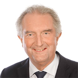
Martin Röcken
Germany
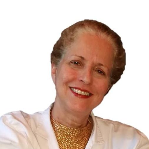
Paola Pasquali
Spain
PRESENTATIONS
Biomarkers that will change clinical practice
12:00 - 12:20 CEST
In recent years there is a translational revolution in atopic dermatitis and other inflammatory skin diseases. With the introduction of multiple new systemic and topical treatments it has become increasingly clear that biomarkers will be useful for adapting the right drug for each patient and for monitoring disease response. This is particularly important as atopic dermatitis, alopecia areata, vitiligo and hidradenitis suppurative among others are heterogeneous diseases, across many disease endotypes and there cannot be a one size fits all treatment. While biopsies are the gold standard for biomarkers, in recent years skin tape stripping showed high accuracy in disease profiling and assessment of biomarkers and responders to treatments. Tape stripping and blood studies are minimally invasive and allow multiple samplings that are often needed in clinical trials and in practice. Skin tape stripping were also shown as an effective way to identify the lung phenotype of patients with atopic comorbidities, and without skin disease.
We will discuss how minimally invasive biomarkers using tape stripping and blood are changing our practice and clinical research in atopic dermatitis and beyond.
Objectives
- Understand utility of biomarkers in atopic dermatitis.
- Learn how biomarkers can be incorporated in clinical trials to objectively assess the skin phenotype.
- Learn how biomarkers can differentiate responders from non responders to treatments and be potentially incorporated into our practice.

Emma Guttman-Yassky
United States
Emma Guttman-Yassky, MD, PhD is the Waldman Professor of Dermatology and Immunology and the health system chair of the department of dermatology at the Icahn School of Medicine at Mount Sinai, New York. She is also the director of the center for excellence in eczema and the laboratory for inflammatory skin diseases and co-director of the Mount Sinai Clinique Skin Aging Center. She earned her M.D. from Sackler School of Medicine at the Tel-Aviv University, and a Ph.D. degree from the Bar-Ilan University, Israel. After her first Board certification in dermatology in Israel, Dr. Guttman moved to the U.S. to pursue a two-year postdoctoral fellowship at The Rockefeller University at the Laboratory for Investigative Dermatology. Upon completion of her fellowship, she completed a second dermatology residency training at Weill-Cornell Medical College, in NY, obtaining her American board certification. Dr. Guttman’s major research focus areas are atopic dermatitis (AD)/eczema, hair loss disorders and skin aging.
Her research made paradigm-shifting discoveries on the immunologic basis of AD/eczema in adults and children, enriching the understanding of this common disorder, opening the door for novel, pathway-specific drugs. Recently, Dr. Guttman extended her research interest to hair loss disorders such as alopecia areata and scarring hair loss, chronic hand eczema, keloids, ichthyosis, and other skin diseases, in which her findings are also translated to novel therapeutics. She has recently made paradigm shifting discoveries linking alopecia areata to the atopic/allergic march, with therapeutic implications. Dr. Guttman co-founded the international eczema council (IEC) and is its past president. Dr. Guttman received many national and international awards (e.g the American Academy of Allergy and Immunology (AAAAI) award for Scientific Innovation and for pioneering discoveries, the Paul Ehrlich award for pioneering discoveries of the European Academy of Allergy and Clinical Immunology (EAACI), the American academy of Dermatology (AAD) Young Investigator award, and many others). She was elected as a member to the American Society for Clinical Investigation (ASCI), and the American Dermatological Society (ADA). She is a co- founder and organizer of one of the most important global meetings in dermatology, the Inflammatory Skin Disease Summit (ISDS), and serves on the editorial board of several high impact journals. Dr. Gutman delivered many keynote and plenary presentations in international and national meetings and authored >420 peer-reviewed publications, including in the New England Journal of Medicine (NEJM), Lancet, Nature, Nature Immunology, Cell, Allergy, and others.
The future of phototherapy in dermatology
12:20 - 12:40 CEST
Phototherapy has entered a paradoxical moment. NB-UVB remains one of dermatology’s safest, most cost-effective and immunologically rational treatments, yet its infrastructure and clinical visibility are being challenged by biologics, JAK inhibitors and other targeted therapies. PUVA, UVA1, and extracorporeal photopheresis are currently used less frequently than before and are now mainly reserved for specific conditions.
This lecture will discuss the future of dermatologic phototherapy in the biologic/JAK era, using current guidelines, expert opinion and recent evidence as a framework. Key themes will include the positioning of NB-UVB, selected UVA approaches and targeted UV treatment for psoriasis, vitiligo, atopic dermatitis, cutaneous T-cell lymphoma and other inflammatory dermatoses. Particular attention will be given to the shift from hospital-based fluorescent-tube systems toward more flexible delivery models, including supervised home phototherapy and emerging UVB-laser and UVB-LED platforms. UVB-LED may enable compact devices, targeted treatment, wavelength-selective output, improved dose control and digital monitoring, but it must be evaluated against established standards. Access to these treatments can be limited by factors like clinic capacity, travel requirements, insurance coverage, staff training, and regulatory oversight.
CONTINUE READING
The key conclusion is that phototherapy continues to be pertinent, particularly when supported by ongoing photodermatology training, and may be effectively integrated with targeted therapies.
Learning objectives
- Explain the current and future role of UV phototherapy in the biologic and JAK inhibitor era, including the continued relevance of NB-UVB, targeted UVB, selected UVA modalities and extracorporeal photopheresis.
- Assess how guidelines, recent evidence and service-delivery challenges are reshaping the use of phototherapy in psoriasis, vitiligo, atopic dermatitis, cutaneous T-cell lymphoma and other inflammatory dermatoses.
- Evaluate emerging models of UV-based phototherapy delivery, including supervised home phototherapy and UVB-LED platforms, with attention to efficacy, safety, equity, governance and evidence gaps.

Lajos Kemény
Hungary
Lajos Kemény is a professor of the Department of Dermatology and Allergology, head of the Skin Research Group, University of Szeged, Hungary. He is an internationally recognized dermatologist, member of the Hungarian Academy of Sciences and member of the Academia Europaea. He served as board member of the EADV, ESDR and UEMS, and he is the Editor-in-Chief of the International Journal of Dermatology. His main research interest is immundermatology and photobiology. He has been awarded by prestigious awards, including Otto Braun-Falco Medal, Kaposi Medal, Certificate of Appreciation for International Leadership Award of the International League of Dermatological Societies, and Scientific Achievement Award of the EADV.
Modern clinical photography in dermatology
12:40 - 13:00 CEST
The essence of dermatology lies in the clinical skill of interpreting and managing the myriad visual appearances of the body’s largest organ. While there is a rich lexicon of words that can be used to describe and classify the vast diversity of the skin in disease and health, the best technology for depicting the skin remains photography–“drawing with light”.
The 21st century development and deployment of universally available digital cameras enabled an explosion in the sheer number of images captured from the skin by health care providers as well as patients; this has forever changed the daily practice of dermatology for the better. Paradoxically however, the ease by which the skin can be recorded almost indiscriminately creates gigabytes, terabytes, and even petabytes of photos of questionable or minimal clinical utility. Furthermore, in the digital era our ability to establish and implement effective policies and procedures to ensure patient safety and improved clinical outcomes has greatly lagged behind the breathtaking speed of technologic advancements.
CONTINUE READING
How do we ensure that clinical photographs are used appropriately? What are the limitations of flat two-dimensional images in clinical practice? Which patients still need to be seen in person? Generative artificial intelligence holds out the promise of propagating synthetic images for education and research, but its role remains to be tested by well-designed studies. Despite all of the technological advances, there is still a long unmet need for dermatologic photographs to be truly representative of the world’s population. Dermatologists also need to dedicate themselves to capturing accurate, informative, and compelling skin images as an essential clinical skill. Just because you have a camera doesn’t mean you are a photographer. Good photography skills are relatively simple to learn, but require a mindful and intentional professional commitment towards self-improvement, practice, and mastery.
Learning objectives:
- To understand how and why photography plays a fundamental role in dermatology.
- To learn the current challenges and pitfalls of current clinical photographic practices and techniques.
- To acquire core clinical skills in photography that will also make you a better dermatologist.

Harvey Lui
Canada
Professor Harvey Lui is an academic dermatologist who specializes in photodermatology, pigmentary skin disorders, skin cancer, lasers, and dermatologic education.
After completing his medical and dermatology training at the University of British Columbia, Prof. Lui completed an advanced fellowship in lasers, photomedicine, and photodynamic therapy at the Massachusetts General Hospital and Harvard Medical School. Prof. Lui was the founding chairman of the only academic dermatology department in Canada.
Prof. Lui has published over 250 peer-reviewed papers, 16 book chapters, and is the co-inventor of seven patents for dermatology. His research group has pioneered technologies for noninvasive optical skin evaluation and also developed the VASI scoring system for assessing vitiligo. He has served as a President of the Canadian Dermatology Association and is an active and honorary member of multiple national and international dermatology societies. In 2015, he was the Secretary-General of the 23rd World Congress of Dermatology in Vancouver, Prof. Lui is also a former President of the International League of Dermatological Societies and is currently working with the WHO on developing a global dermatology image reference gallery based on ICD-11 (www.ILDSDermGallery.org).
Plenary lectures B
Room: Hall A
Date: Thursday, 01 October, 12:00 - 13:00 CEST
CHAIRS
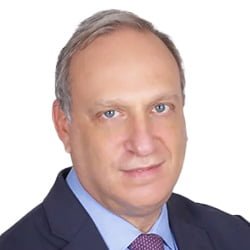
Alexander Stratigos
Greece

Cristina Has
Germany
PRESENTATIONS
Tumor microenvironment in skin cancer
12:00 - 12:20 CEST
The tumor microenvironment (TME) is a critical determinant of skin cancer initiation, progression, and therapeutic response. Immune cells, fibroblasts, endothelial cells, the extracellular matrix, the microbiome, and the complement system interact with malignant keratinocytes and melanocytes to shape skin cancer. Beyond its classical role in innate immunity, complement influences inflammation, tumor-cell survival, angiogenesis, stromal remodeling, and immune surveillance.
In UV-exposed skin, chronic inflammation, DNA damage, and immunosuppression create a permissive environment for tumor progression and clonal selection. Recent single-cell and spatial transcriptomic studies have shown how skin cancers co-opt wound-healing and stromal-remodeling programs to promote growth, invasion, and immune evasion. Complement activation may amplify inflammatory signaling and regulate interactions among tumor, immune, and stromal cells, whereas dysregulated complement activity may promote immunosuppression and tumor persistence.
CONTINUE READING
The immune contexture of the TME includes T cells, myeloid-derived suppressor cells, tumor-associated macrophages, dendritic-cell subsets, and complement-mediated pathways. Together, these components influence responses to immune checkpoint blockade and other immunotherapies. The balance between complement-mediated inflammation and immune suppression may be particularly important for treatment response. PD-1/PD-L1, CTLA-4, and emerging checkpoints function within the distinctive immunologic landscape of the skin, yet some tumors remain immunologically “cold” despite a high mutational burden. Complement components and their receptors may contribute by regulating immune-cell recruitment, activation, and exhaustion.
Non-immune components—including cancer-associated fibroblasts, perineural niches, lymphatic remodeling, and vascular alterations—promote metastasis and resistance to targeted and immune therapies. Complement signaling may intersect with these processes through effects on fibroblast activation, extracellular-matrix remodeling, angiogenesis, and vascular permeability.
Translational strategies for reprogramming the skin cancer microenvironment include rational immunotherapy combinations, stromal-targeting approaches, complement-directed interventions, and treatments tailored to high-risk states such as immunosuppression and field cancerization. Defining complement activity within the TME may provide biomarkers for patient stratification and identify new therapeutic targets, informing next-generation clinical trials aimed at improving skin cancer control, treatment response, and prevention.
Learning objectives:
By the end of the presentation, participants will be able to
- Describe the cellular, stromal, extracellular, microbial, and complement-system components of the tumor microenvironment in skin cancer.
- Explain how immune contexture, complement activity, chronic inflammation, wound-healing programs, and stromal remodeling contribute to tumor progression, immune evasion, metastasis, and treatment resistance.
- Evaluate the potential of complement-directed therapies, stromal-targeting strategies, and tumor-microenvironment biomarkers to improve patient stratification and responses to immunotherapy.

Veli-Matti Kähäri
Finland
Dr. Veli-Matti Kähäri, MD, PhD is Professor and Chief Physician, Department of Dermatology, University of Turku and Turku University Hospital (2005-) and Head of the Institute of Clinical Medicine, University of Turku, Turku, Finland (2022-). Dr Kähäri earned his MD in 1984 and PhD in 1987 at University of Turku, and became specialist in Dermatology and venereology in 1995. He served as postdoctoral fellow (1988-89) and as Research Assistant Professor (1989-91) at the Department of Dermatology of Thomas Jefferson University, Philadelphia, PA, USA. He has extensive experience on translational research in skin cancer, wound repair, and dermal fibrosis. His current research focus is molecular mechanisms and biomarkers of skin cancer progression.
Dr. Kähäri is a member of Finnish Academy of Science and Letters (2009-). He is the author of 180 original publications and 77 reviews and book chapters. His articles have been cited 20270 times and his H-Index is 76. He has served as board member of European Society for Dermatological Research, European Dermatology Forum and UEMS Dermato-Venereology Section and is currently member of the board of EADV.
New algorithms for mananging high-risk skin cancer patients
12:20 - 12:40 CEST
Skin cancer is the most common malignancy worldwide, and its incidence continues to rise owing to population aging, cumulative ultraviolet (UV) exposure, increasing numbers of immunosuppressed patients, and improved diagnostic awareness. Among keratinocyte carcinomas, cutaneous squamous cell carcinoma (CSCC), the focus of this presentation, encompasses a highly heterogeneous spectrum of disease, ranging from precancerous lesions such as actinic keratosis and squamous cell carcinoma in situ (Bowen’s disease) to invasive tumors, which can be stratified into low- and high-risk categories, and ultimately to advanced disease, including locally advanced and metastatic CSCC. Although most CSCCs are effectively treated with surgery, approximately 3–5% of patients develop locally advanced or metastatic disease, accounting for the vast majority of disease-specific deaths. High-risk invasive and advanced CSCC are characterized by clinicopathological features associated with an increased risk of local recurrence, nodal metastasis, locoregional recurrence, and disease-specific mortality.
CONTINUE READING
Tumor-related characteristics remain the cornerstone of risk stratification. Larger tumor diameter, specific anatomical locations such as the ear, lip, temple, and scalp, increasing depth of invasion beyond the subcutaneous tissue, poor histological differentiation, lymphovascular invasion, and especially perineural invasion are consistently associated with a significantly higher risk of recurrence and metastatic spread. Host-related factors also contribute substantially to prognosis. Immunosuppressed patients, including solid-organ transplant recipients, individuals with hematologic malignancies, and those receiving long-term immunosuppressive therapy, develop more aggressive tumors with higher recurrence rates and poorer survival. Nevertheless, currently available staging systems have several limitations, and there is still no consensus regarding the optimal risk stratification model for routine clinical practice.
Although the 8th edition of the American Joint Committee on Cancer (AJCC-8) staging system remains the international standard for many cutaneous malignancies, it was developed exclusively for CSCC of the head and neck and does not incorporate several important pathological and patient-related variables, including tumor differentiation and immune status. Alternative staging systems, such as the Brigham and Women’s Hospital (BWH), improve prognostic accuracy by incorporating additional pathological variables associated with metastatic potential. In particular, BWH T2b and T3 tumors account for the majority of nodal metastases and disease-specific deaths despite representing only a minority of all primary tumors. Nevertheless, no single classification fully captures the biological heterogeneity of high-risk CSCC. More recently, multidimensional prognostic models integrating clinicopathological variables, molecular biomarkers, and artificial intelligence-assisted image analysis have shown considerable promise in providing more refined risk estimates and improved prognostic discrimination, particularly for clinically relevant outcomes such as metastasis and disease-specific mortality, while enabling individualized risk assessment based on key clinical and histopathological variables.
Therapeutic strategies have also evolved substantially over the last years. The management of high-risk CSCC increasingly relies on a multidisciplinary approach involving dermatologists, dermatologic surgeons, surgical oncologists, plastic surgeons, radiation oncologists, medical oncologists, pathologists, radiologists, and, when appropriate, head and neck surgeons. Such multidisciplinary evaluation allows optimal integration of surgery, radiotherapy, and systemic treatment according to tumor stage and patient characteristics.
Surgical excision remains the first-line treatment for most patients. Complete histological margin assessment is critical, as inadequate excision significantly increases the risk of local recurrence and may compromise subsequent therapeutic options. Whenever feasible, Mohs micrographic surgery is considered the treatment of choice for recurrent tumors, lesions with poorly defined clinical margins, tumors located in anatomically critical areas, and lesions exhibiting aggressive histopathological characteristics.
Radiotherapy continues to play an important role in selected patients, particularly those who are poor surgical candidates or present with positive margins, extensive perineural invasion, or regional nodal involvement. Adjuvant radiotherapy may improve local control in selected high-risk cases, although its impact on overall survival remains less clearly established.
A remarkable therapeutic advance in recent years has been the introduction of immune checkpoint inhibitors targeting the PD-1 pathway. CSCC is characterized by one of the highest tumor mutational burdens among human malignancies as a consequence of chronic ultraviolet radiation exposure, making it particularly immunogenic and therefore highly responsive to immune checkpoint blockade.
Cemiplimab was the first PD-1 inhibitor approved for patients with advanced CSCC who are not candidates for curative surgery or radiotherapy. In the pivotal EMPOWER-CSCC-1 trial, cemiplimab achieved objective response rates of approximately 44–50%, with complete responses observed in nearly 17% of patients following longer follow-up. Importantly, clinical benefit was maintained beyond two years in the the majority of responding patients. Furthermore, long-term analyses demonstrated encouraging overall survival outcomes together with an acceptable safety profile, establishing PD-1 blockade as the standard first-line systemic treatment for unresectable locally advanced and metastatic CSCC. Pembrolizumab has subsequently expanded the therapeutic armamentarium, showing comparable efficacy and durable responses in patients with recurrent or metastatic disease, thereby providing an additional therapeutic option in selected clinical settings.
The role of immunotherapy has progressively expanded from advanced disease to the perioperative setting. Adjuvant immunotherapy is emerging as a new standard of care for patients with high-risk resected CSCC. In the randomized phase III C-POST trial, adjuvant cemiplimab reduced the risk of disease recurrence or death by approximately 68% compared with placebo (HR 0.32), resulting in a clinically meaningful improvement in disease-free survival. These findings represent the first robust evidence supporting adjuvant systemic therapy in resected high-risk CSCC and are expected to substantially modify future treatment algorithms for selected patients.
Even more remarkable are the results obtained with neoadjuvant immunotherapy, which represents a paradigm shift in the management of resectable, locally advanced CSCC. In the phase II study evaluating neoadjuvant cemiplimab, 51% of patients achieved a pathological complete response (pCR), while an additional 13% achieved a major pathological response (MPR), resulting in an overall pathological response rate of 64%. Interestingly, pathological responses substantially exceeded radiological complete responses, highlighting the limitations of imaging in accurately assessing residual viable tumor after immunotherapy. These impressive pathological response rates suggest that neoadjuvant PD-1 blockade can effectively downstage tumors, facilitate less extensive surgical procedures, reduce surgical morbidity, and potentially improve long-term oncological outcomes. Ongoing randomized studies with longer follow-up will determine whether these early pathological responses ultimately translate into improved disease-free and overall survival while also identifying predictive biomarkers capable of selecting patients most likely to benefit from perioperative immunotherapy.
Alongside systemic treatment, intralesional therapy represents an attractive therapeutic option for selected patients with CSCC, particularly elderly or frail individuals for whom surgery or systemic immunotherapy may not be feasible. Intralesional immunotherapy aims not only to induce direct tumor regression but also to stimulate systemic antitumor immune responses capable of controlling both injected and non-injected lesions through activation of adaptive immunity. Several innovative strategies are currently under clinical investigation, including talimogene laherparepvec (T-VEC), RP1, Daromun (L19IL2/L19TNF), STING agonists, RNA-based immune stimulators, and other locally delivered immunomodulatory agents. Although available evidence is still largely derived from early-phase clinical trials, these approaches have demonstrated encouraging local response rates together with favorable safety profiles, raising the possibility that intralesional immunotherapy may become an important component of multimodal treatment strategies for selected patients with localized or oligometastatic disease.
In conclusion, the management of high-risk CSCC has evolved from a predominantly stage-based approach to a dynamic, individualized strategy that integrates clinicopathological characteristics, multidisciplinary evaluation, and increasingly effective systemic therapies to guide clinical decision-making. The introduction of immune checkpoint inhibitors has fundamentally transformed the therapeutic landscape, not only improving survival in advanced disease but also opening new opportunities in both the neoadjuvant and adjuvant settings. At the same time, advances in molecular pathology, digital imaging, artificial intelligence, and precision oncology are expected to further refine prognostic assessment and therapeutic selection. Future risk stratification algorithms will likely combine traditional histopathological parameters with molecular biomarkers and computational prediction models, enabling earlier identification of patients at highest risk, more personalized treatment selection, avoidance of unnecessary interventions, and ultimately improved survival, functional outcomes, and quality of life.
Learning objectives:
- Risk stratification of patients with high-risk basal cell carcinoma and squamous cell carcinoma
- Evidence-based management algorithms according to the latest international guidelines
- Emerging therapeutic strategies and future perspectives

Ketty Peris
Italy
Professor Ketty Peris is Full Professor and Chair of the Department of Dermatology at the Università Cattolica del Sacro Cuore and Head of the School of Specialization in Dermatology and Venereology, Rome, Italy. She is an internationally recognized dermatologist and clinician-scientist with more than three decades of experience in skin cancer, dermato-oncology, dermoscopy, psoriasis, and inflammatory skin diseases.
After graduating summa cum laude in Medicine from Sapienza University of Rome and completing her specialization in Dermatology with highest honors, Professor Peris undertook postgraduate training in pathology at the University of Munich, Germany, and clinical dermatology at the University of Graz, Austria. She began her academic career at the University of L’Aquila, where she progressed from Assistant Professor to Full Professor and served as Head of the Department of Dermatology and Director of the School of Specialization in Dermatology and Venereology before joining the Catholic University of the Sacred Heart in 2014.
Professor Peris has played a leading role in national and international dermatological societies. She served as President of the Italian Society of Medical, Surgical, Aesthetic Dermatology and Sexually Transmitted Diseases (SIDeMaST), is General Secretary of the European Association of Dermato-Oncology (EADO), General Secretary of the 24th World Congress of Dermatology (Milan, 2019), and currently serves on the Executive Committee of the International League of Dermatological Societies (ILDS). She is also a member of the Board of Directors of Università Cattolica del Sacro Cuore.
Her research has focused on the early diagnosis and prevention of melanoma and non-melanoma skin cancer, dermoscopy, translational dermato-oncology, and immune-mediated diseases as psoriasis and atopic dermatitis. She has coordinated and participated in numerous nationally funded research projects and clinical studies and has extensive experience in Good Clinical Practice (GCP)-compliant clinical research.
Professor Peris has authored 735 peer-reviewed scientific publications and has delivered more than 500 invited lectures at national and international scientific meetings.
Social media for doctors. From viral posts to legal warnings
12:40 - 13:00 CEST
Social media has transformed how patients obtain information about skin and hair disorders. It has also created a paradox: scientifically unsupported claims from influencers can reach millions of users, while healthcare professionals attempting to provide evidence-based information may face significant ethical, regulatory and legal risks.
This lecture will examine why dermatology is particularly susceptible to viral content. Visible outcomes, before-and-after images, emotionally charged conditions and simplified treatment rankings can make anecdotal evidence appear more persuasive than clinical data. Recent research on sunscreen and hair-loss content illustrates how low-quality or misleading information can achieve disproportionate engagement.
CONTINUE READING
The presentation will then explore the boundary between independent scientific information and pharmaceutical advertising. Although European countries share a common legal framework prohibiting public advertising of prescription-only and unauthorised medicines, the interpretation and enforcement of these rules differ between jurisdictions. Relevant approaches from selected specific countries of the European Union will be compared. Particular attention will be paid to active ingredients, off-label uses, investigational treatments, compounded formulations, patient testimonials, before-and-after images and professional-only communications.
The speaker will share his personal experience of receiving an administrative requirement from a Spanish health authority following a third-party complaint concerning educational content published on a website and several social-media platforms. The requirement demanded the review and removal of allegedly unlawful content within ten working days and cited potential sanctions applicable to very serious infringements. This case demonstrates how scientific intent alone may not determine whether online content is regarded as information or promotion.
Finally, participants will be introduced to a practical “Five-P” framework—Purpose, Product, Public, Payment and Presentation—and a green–amber–red risk model to help healthcare professionals assess content before publication. The aim is not to discourage doctors from communicating online, but to promote evidence-based, transparent and legally responsible participation in the digital public conversation.
Learning objectives
At the end of this lecture, participants will be able to:
- Distinguish independent scientific or disease-awareness information from content that may be classified as pharmaceutical advertising on public social-media platforms.
- Identify the principal legal and professional risks associated with discussing prescription-only, off-label, investigational or compounded treatments online across different European jurisdictions.
- Apply the Five-P framework and the green–amber–red risk model to assess and improve the scientific balance, transparency and legal safety of their own social-media content before publication.

Sergio Vaño-Galván
Spain
He is the former President of the Spanish Hair Research Society (2019-2023). Dr. Vañó-Galván leads an active research group in the study of different forms of alopecia (#TricoHRC). Dr. Vañó-Galván has published over 370 scientific articles in Pubmed (h-index of 40) and is a regular guest speaker at international congresses of hair research.
He is the President of the European Hair Research Society (2026-2029) and he is an American Hair Research Society member. He is the Director of the International Master’s Degree in Hair Disorders and Hair Transplantation of the University of Alcalá since 2016.
Future President of the World Congress for Hair Research (Madrid, 2028)
Dr. Vañó-Galván also works as a Section Editor in the Journal of the European Academy of Dermatology (JEADV) and is an active user of social networks
Plenary lectures C
Room: Hall A
Date: Saurday, 03 October, 12:00 - 13:00 CEST
CHAIRS

Luca Borradori
Switzerland
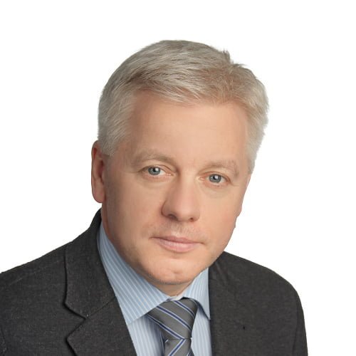
Rolland Gyulai
Hungary
PRESENTATIONS
RTF plenary lecture: Personalized treatment for skin IMIDs
12:00 - 12:20 CEST
The treatment of immune-mediated inflammatory diseases (IMIDs) of the skin has been revolutionized by biologics and small molecules targeting specific immune pathways. Psoriasis, atopic dermatitis, lichen planus and lupus are increasingly understood through the dominant immune pathways driving inflammation, including Th17, Th2, Th1 and type I interferon responses. However, the expanding therapeutic armamentarium raises a major clinical challenge: how can we select the right targeted treatment for the individual patient?
In this lecture, I will present a precision medicine approach in which treatment decisions are guided by the molecular profile of a patient’s disease.
CONTINUE READING
Using transcriptomic analysis of skin biopsies, we have defined immune modules representing Th17, Th2, Th1, type I interferon, neutrophilic, macrophagic and eosinophilic inflammation and constructed a molecular immune cartography of inflammatory skin diseases. A digital platform allows individual patient samples to be positioned within this reference map and their dominant immune modules to be quantified. This approach has important implications for both diagnosis and treatment. Molecular mapping can distinguish clinically overlapping inflammatory dermatoses, including difficult presentations such as erythroderma and palmoplantar disease, and thereby prevent inappropriate treatment resulting from diagnostic misclassification. More importantly, identification of the dominant immune module provides a rationale for matching each patient with the targeted therapy most likely to inhibit the pathway driving their disease. Molecular profiling can also help us understand why targeted treatments fail. In most non-responding patients, the pathway targeted by therapy does not correspond to the dominant immune pathway detected in the lesion. This may result from an incorrect diagnosis or from an immune shift, in which therapeutic pressure leads to the emergence of an alternative dominant pathway. Such shifts can explain acquired resistance and paradoxical reactions and provide a biological rationale for adapting or broadening therapy.
Finally, identification of shared immune modules across different diseases creates opportunities for rational repurposing of existing targeted therapies beyond their established indications.
Together, these findings introduce a new paradigm for the personalized management of skin IMIDs: moving beyond treatment based solely on disease labels toward treatment based on the molecular architecture of an individual patient’s disease. Integrating molecular diagnosis, immune pathway identification and targeted therapy has the potential to improve treatment selection, explain therapeutic failure and bring precision medicine into routine dermatological care.
Learning objectives:
- Understand how molecular immune profiling can identify the dominant inflammatory pathways underlying skin IMIDs.
- Apply immune-module profiling to improve diagnosis and personalize the selection of targeted therapies for individual patients.
- Recognize how diagnostic mismatch, immune shifts and paradoxical reactions contribute to treatment failure and how molecular profiling can guide therapeutic adaptation and drug repurposing.

Michel Gilliet
Switzerland
Michel Gilliet, M.D., is Head of the Dermatology Department at the University Hospital CHUV in Lausanne, Switzerland. He completed his dermatology training at the University of Zurich and received research training in immunology at the DNAX Research Institute in Palo Alto, California. In 2004, Dr. Gilliet was recruited by MD Anderson Cancer Center in Houston, Texas, where he held clinical appointments in Dermatology, as well as basic and translational science appointments in Melanoma Oncology and Immunology.
In 2010, Dr. Gilliet returned to Switzerland as a Full Professor and Head of the Dermatology Department in Lausanne. His research primarily focuses on immune responses in inflammatory skin diseases. His laboratory has notably uncovered mechanisms by which dendritic cells initiate and sustain skin inflammation by sensing DNA in complex with antimicrobial peptides. These groundbreaking studies have led to a paradigm shift in understanding how inflammation is regulated in injured skin and in diseases such as psoriasis and lupus erythematosus.
More recently, his lab has expanded its research to include the application of molecular omics technologies in clinical dermatology practice, providing a new framework for diagnosing and treating skin diseases.
Dr. Gilliet’s research has led to over 150 publications and garnered 32,000 citations, with his work regularly featured in prestigious journals such as Nature, Science, Nature Immunology, Nature Medicine, and Nature Communications.
Dr. Gilliet has served as President of the European Society for Dermatological Research (ESDR) and is currently President of the Skin Science Foundation. He also serves on the Board of the European Academy of Dermatology and Venereology (EADV), where he chairs the Scientific Program. In recognition of his contributions to medical research, he received the Cloetta Prize, Switzerland’s most prestigious medical research award, in 2016. In 2023, he was elected to the National Academy of Sciences in Germany.
ESDR plenary lecture - Genetic mechanisms in eczema: relevance to the patient and clinician
12:20 - 12:40 CEST
This talk will provide an overview of the molecular and genetic mechanisms which lead to atopic eczema and related skin and systemic disorders. The presentation will highlight the importance of mechanistic understanding to improve clinical management of skin inflammation and a perspective for future targeted therapies.
Learning objectives
- Describe the role of genetic predisposition in the common inflammatory skin disease atopic eczema.
- Understand how molecular mechanisms have guided the development of targeted anti-inflammatory therapies.
- Discuss future needs and opportunities towards personalised medicine for patients with atopic eczema.

Sara Brown
United Kingdom
Sara Brown is a clinical academic dermatologist, combining her specialist interest in atopic eczema with research into the molecular and genetic mechanisms of skin barrier function and inflammatory skin disease. Sara trained in Dermatology in Newcastle-upon-Tyne, UK, with Prof Nick Reynolds and Peter Farr, followed by specialist training in Paediatric Dermatology and Genodermatoses in Dublin, Ireland, with Prof Alan Irvine and Dr Rosemary Watson. Sara holds an MD and PhD in Human Genetics; her early work focussed on the role of filaggrin in atopic skin disease, in the laboratory of Prof Irwin Mclean, Dundee, Scotland. Sara now holds the Grant Chair of Dermatology at the University of Edinburgh.
The misfiring alarm: Lessons from fibromyalgia for stress-driven skin disease
12:40 - 13:00 CEST
What if the skin lesion is not the disease — but its most visible consequence? This lecture invites dermatologists to look upstream: to the central alarm system whose dysregulation may be driving what they see and treat at the surface. Drawing on the FITSS model (Fibromyalgia: Imbalance of Threat and Soothing Systems), published in Nature Reviews Rheumatology (2023), this talk proposes that the central nervous system mechanisms underlying fibromyalgia — a chronically misfiring alarm sustained by imbalance between threat-detection and soothing-safety systems, mediated by the Salience Network — may illuminate the pathophysiology of stress-driven skin conditions in ways that current inflammation-centred models do not.
In fibromyalgia, the peripheral tissues are essentially intact. The pain is real, but its generator is central: a nervous system locked in a state of threat, unable to access the soothing and safety signals that would restore equilibrium. The skin, by contrast, displays unmistakable peripheral pathology — yet the evidence consistently shows that stress precipitates, maintains, and amplifies conditions such as psoriasis, atopic dermatitis, chronic spontaneous urticaria, alopecia areata, and chronic itch. The central alarm system, it appears, is not a bystander.
CONTINUE READING
The therapeutic implication is direct: targeting the alarm — through interventions that restore the balance between threat and soothing systems — may be as important as suppressing local inflammation. Emerging evidence from mindfulness-based interventions, compassion-focused approaches, and the neuroscience of care context in itch perception begins to sketch this complementary paradigm. A further possibility, raised as a hypothesis rather than a claim, is that the skin — unlike muscle in fibromyalgia — may possess mechanisms for autonomous peripheral amplification of the central alarm signal. Whether this represents a meaningful therapeutic target is a question this lecture will pose to the dermatological community.
This is not a lecture about fibromyalgia. It is a lecture about what a misfiring alarm looks like — and what it might take to silence it.
Using transcriptomic analysis of skin biopsies, we have defined immune modules representing Th17, Th2, Th1, type I interferon, neutrophilic, macrophagic and eosinophilic inflammation and constructed a molecular immune cartography of inflammatory skin diseases. A digital platform allows individual patient samples to be positioned within this reference map and their dominant immune modules to be quantified. This approach has important implications for both diagnosis and treatment. Molecular mapping can distinguish clinically overlapping inflammatory dermatoses, including difficult presentations such as erythroderma and palmoplantar disease, and thereby prevent inappropriate treatment resulting from diagnostic misclassification. More importantly, identification of the dominant immune module provides a rationale for matching each patient with the targeted therapy most likely to inhibit the pathway driving their disease. Molecular profiling can also help us understand why targeted treatments fail. In most non-responding patients, the pathway targeted by therapy does not correspond to the dominant immune pathway detected in the lesion. This may result from an incorrect diagnosis or from an immune shift, in which therapeutic pressure leads to the emergence of an alternative dominant pathway. Such shifts can explain acquired resistance and paradoxical reactions and provide a biological rationale for adapting or broadening therapy.
Finally, identification of shared immune modules across different diseases creates opportunities for rational repurposing of existing targeted therapies beyond their established indications.
Together, these findings introduce a new paradigm for the personalized management of skin IMIDs: moving beyond treatment based solely on disease labels toward treatment based on the molecular architecture of an individual patient’s disease. Integrating molecular diagnosis, immune pathway identification and targeted therapy has the potential to improve treatment selection, explain therapeutic failure and bring precision medicine into routine dermatological care.
Learning objectives:
- Understand how molecular immune profiling can identify the dominant inflammatory pathways underlying skin IMIDs.
- Apply immune-module profiling to improve diagnosis and personalize the selection of targeted therapies for individual patients.
- Recognize how diagnostic mismatch, immune shifts and paradoxical reactions contribute to treatment failure and how molecular profiling can guide therapeutic adaptation and drug repurposing.

José Antonio Pereira da Silva
Portugal
He has published over 340 papers in National and International peer-reviewed journal (HI 58, ORCID 0000-0002-2782-6780) and currently serves in the Editorial Board of several scientific journals in the area of Rheumatology. His current research interests are especially focused in Rheumatoid arthritis, Osteoporosis, Systemic sclerosis, Fibromyalgia and the psychological domains of rheumatic diseases.
He especially prides himself on being a medical educator and a practicing clinician dedicated to translating research into the best possible practice to the benefit of patients, thus pursuing the moto he adopted for his department: “Promoting happiness through excellence of care”.