EADV Congress 2025 - Scientific session takeaways
Our medical writers prepared daily round-ups of selected scientific sessions.
Plenary session with keynote lecture
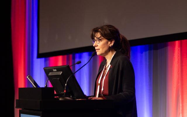
Live like a sloth, think like an octopus: Lessons from nature’s greatest survivors
We were honoured to be joined by Lucy Cooke for today’s keynote lecture. Lucy is a New York Times best-selling author, National Geographic explorer, TED talker, and award-winning documentary producer and presenter. She also holds a Master’s in zoology from New College, Oxford, where she studied evolution and animal behaviour.
In her fascinating talk, Lucy explored how unconscious bias has shaped our understanding of the natural world. Take sloths, for instance: long dismissed as lazy, they actually emerge as remarkable champions of sustainability, perfectly attuned to their environment. Or consider the silverback gorilla, whose soft and nurturing side contrasts sharply with the enduring myth of the aggressive alpha male.

As Lucy explained, while we may be living in extraordinarily uncertain times, nature holds many of the answers. Through striking photos, compelling videos, and insightful anecdotes from her time in the field, she introduced us to some of nature’s greatest survivors – revealed in a new light, beyond the bias.
Psoriasis
Understanding endotypes
Prof. Denis Jullien (Lyon, France) first reminded the audience that psoriasis is a complex heterogeneous condition comprised of distinct endotypes of clinical relevance – the focus of his presentation.
Identification of endotypes aims to rationalise disease diversity by stratifying patients into distinct disease subgroups based on shared molecular characteristics, genetic markers (e.g. interleukin [IL]-36 and interferon [IFN]-1 pathways and HLA-C*06:02 status) and treatment responses. This represents a paradigm shift from a phenotype-based approach to mechanism-based disease classification and stratified treatment.
Endotypes represent a paradigm shift from phenotype- to mechanism-based disease classification.
Prof. Denis Jullien
Recent research that has enabled the integration of genetic, molecular, cellular, and clinical data has revealed multiple overlapping endotype systems, with each providing unique insights into disease heterogeneity.As a result, the clinical translation of each endotype holds immense promise for precision medicine in treating psoriasis.
Furthermore, emerging biomarker panels and molecular signatures are being developed for treatment selection and will likely involve the use of artificial intelligence.
Although important advances have already been made, significant challenges still remain, including validation of biomarkers, standardisation of measurement protocols and cost-effective implementation intodaily clinical practice.
Towards a cure: Hit early, hit hard
Dr. Andrew Blauvelt M.D. (Portland, USA) provided his insights into an early intervention approach with high-dose biologics for psoriasis. Although disease remission has traditionally been the mainstay of treatment, a ‘cure’ for psoriasis has been proposed that restores immune homeostasis using a combination approach alongside personalised medicine. Using this approach, early intervention with high-dose biologics, advanced therapeutics and lifestyle intervention are key.
Hitting early and hard may be the best future therapeutic strategy, leading to prolonged remission and even a cure in some patients.
Dr. Andrew Blauvelt M.D.
Treating patients with short-duration disease (e.g. less than 1–2 years) with biologics has led to improved efficacy and ability to control disease over time, with complete skin clearance (psoriasis area and severity index [PASI] 100 scores) without recurrence, termed ‘super responders.’ Furthermore, research has shown that early intervention with IL-23 inhibitors in patients with short disease duration can lead to earlier normalisation of CD8+ tissue-resident memory cells (responsible for recurrence at the same disease site) and unprecedented PASI 100 scores. Importantly, knockout data suggest that high induction doses with IL-23 inhibitors are safe, offering the ability to increase clearance rates and remission times, as an alternative to established dosing patterns.
Combination therapies are currently exploring the ‘hit-hard, hit early’ approach and these results are eagerly awaited
Oral treatments
In his presentation, Prof. Richard B. Warren (Manchester, UK), first asked the audience if oral treatments are needed in the advent of newer biologics, before presenting situations where they may be warranted, including patients with a needle phobia; as a low-cost option; as a faster route to prescription/improved transport; or in case of immunogenicity concerns.
Phosphodiesterase-4 (PDE-4) and tyrosine kinase-2 (TYK-2) inhibitors represent available oral options for psoriasis which have been developed over the last decade. He noted that while efficacy with PDE-4 inhibitors remains modest, safety can be an advantage.
The oral peptide data are very strong and are setting the standard for oral therapies from both efficacy and safety perspectives.
Prof. Richard B. Warren
Advances using TYK-2 inhibitors allow therapy to be more targeted. Phase 2 data with second-generation compounds show competitive levels of efficacy and long-term retention of response; higher levels of efficacy are anticipated in phase 3 studies. The safety profile is reassuring but minimal screening and monitoring are recommended.
He then discussed the role of cytokines, in which the treatment landscape is changing, with oral molecules or peptide-inhibiting cytokines being developed for psoriasis. These include icotrokinra, a selective IL-23 inhibitor and upadacitinib, a janus kinase [JAK] inhibitor, notably in studies that included adolescents (>12 years) and high-impact site involvement.
These compounds may be the start of a new psoriasis treatment paradigm, with IL-23 peptide treatments setting the standard for oral therapies from both efficacy and safety perspectives. Newer data are eagerly awaited and may break down barriers in treating more ‘moderate’ disease.
Diet and psoriasis
As patients often ask clinicians, and clinicians need to know whether dietary advice should be part of management, Prof. Wendy Hall (London, UK) outlined the importance of diet in managing psoriasis.
Psoriasis is a chronic inflammatory condition with inherent links to obesity, cardiometabolic disease, and mental health. High intake of saturated fat, sugar, processed meat, salt, and alcohol can drive pro-inflammatory pathways, while diets rich in fruits, vegetables, oily fish, unsaturated oils, polyphenols, and fibre have anti-inflammatory effects. Vitamin D, probiotics, and prebiotics are also potential modulators of immune pathways.
Mediterranean and anti-inflammatory dietary patterns are promising but not yet guideline-level evidence.
Prof. Wendy Hall
Weight reduction, especially in individuals with obesity, improves severity and treatment response. Guidelines focus on weight; however, reduction is rarely maintained over time, and not all patients have overweight/obesity.
Current evidence on diet quality suggests the diets with anti-inflammatory properties are important; however, the evidence base is still emerging. Anti-inflammatory diets such as the Mediterranean diet may be beneficial, but robust well-powered RCTs that prioritise plant-based, Mediterranean, and time-restricted eating [fasting ≥10 hours/day] diets are needed.
There is also a need to consider potential heterogeneity of psoriasis phenotypes and responses, and patient preference/personalisation should be used to guide clinical advice.
Key Takeaways:
- Targeted cytokine therapies are advancing efficacy and convenience across inflammatory diseases, with oral agents and multi-cytokine blockers offering new options beyond injectables
- Strategic immune modulation that considers timing, combinations, and upstream targeting is key to durable outcomes in conditions like alopecia areata, vitiligo, and autoimmune skin diseases
JAK inhibitors
Approved drugs and their indications
Prof. Kamran Ghoreschi (Berlin, Germany) provided an update on JAK and TYK2 inhibitors in dermatology. He highlighted the progress of topical formulations, which have shown durable efficacy with minimal systemic absorption in vitiligo and hand eczema. For systemic therapy, he noted long-term efficacy in atopic dermatitis and alopecia areata, with real-world findings confirming sustained hair regrowth.
Phase 3 data in giant cell arteritis demonstrated reduced flare rates, while safety analyses covering >27,000 patient-years revealed no new signals.
Prof. Ghoreschi also pointed to biomarker studies linking immune pathway modulation with clinical regrowth in alopecia and four-year psoriasis extension data confirming durable PASI responses.
Off-label use
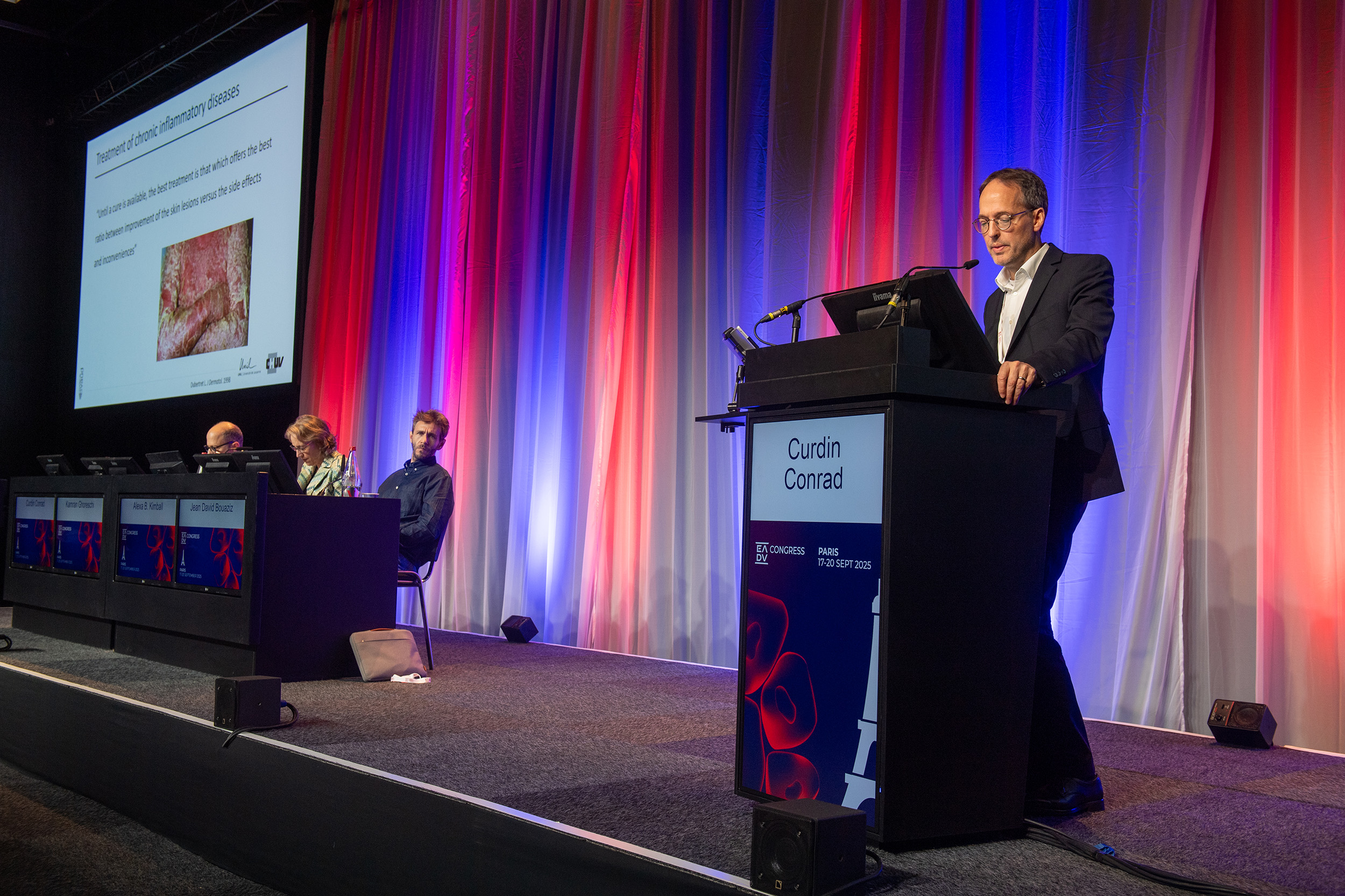
Prof. Curdin Conrad (Lausanne, Switzerland) expanding on the role of JAK/TYK2 inhibition, emphasised their capacity to modulate multiple inflammatory pathways simultaneously.
Unlike biologics that act on single cytokines, these inhibitors target converging Th1, Th2, and Th17 circuits, making them well suited to conditions with overlapping immune signatures. Molecular profiling has revealed distinct patterns across diseases and by interrupting these signalling hubs, JAK/TYK2 blockade dampens interferon-driven inflammation, reduces lymphocyte effector function, and restores tissue homeostasis.
JAK inhibition provides an intriguing therapeutic option for a growing list of skin conditions.
Prof. Denis Jullien
Short and long term monitoring
Prof. Jean-David Bouaziz (Paris, France) opened his talk by asking, “Are JAK inhibitors the new steroids?” He highlighted their rapid onset, broad immunologic activity, and oral/topical availability as advantages, while noting that safety remains a central concern. The 2021 FDA black box warning, based on rheumatoid arthritis data, has influenced prescribing practices, even among younger dermatology patients. However, real-world data suggest most adverse events are mild.
Prof. Bouaziz stressed the importance of comprehensive baseline assessment, including cardiovascular, infection, and malignancy risk, alongside ongoing monitoring of lipids and tuberculosis status.
Dermatologists should leverage these versatile agents judiciously, considering patient-specific factors and maintaining vigilant monitoring to optimise outcomes while minimising risks.
Prof. Denis Jullien
Pipeline of systemic and topical JAKs
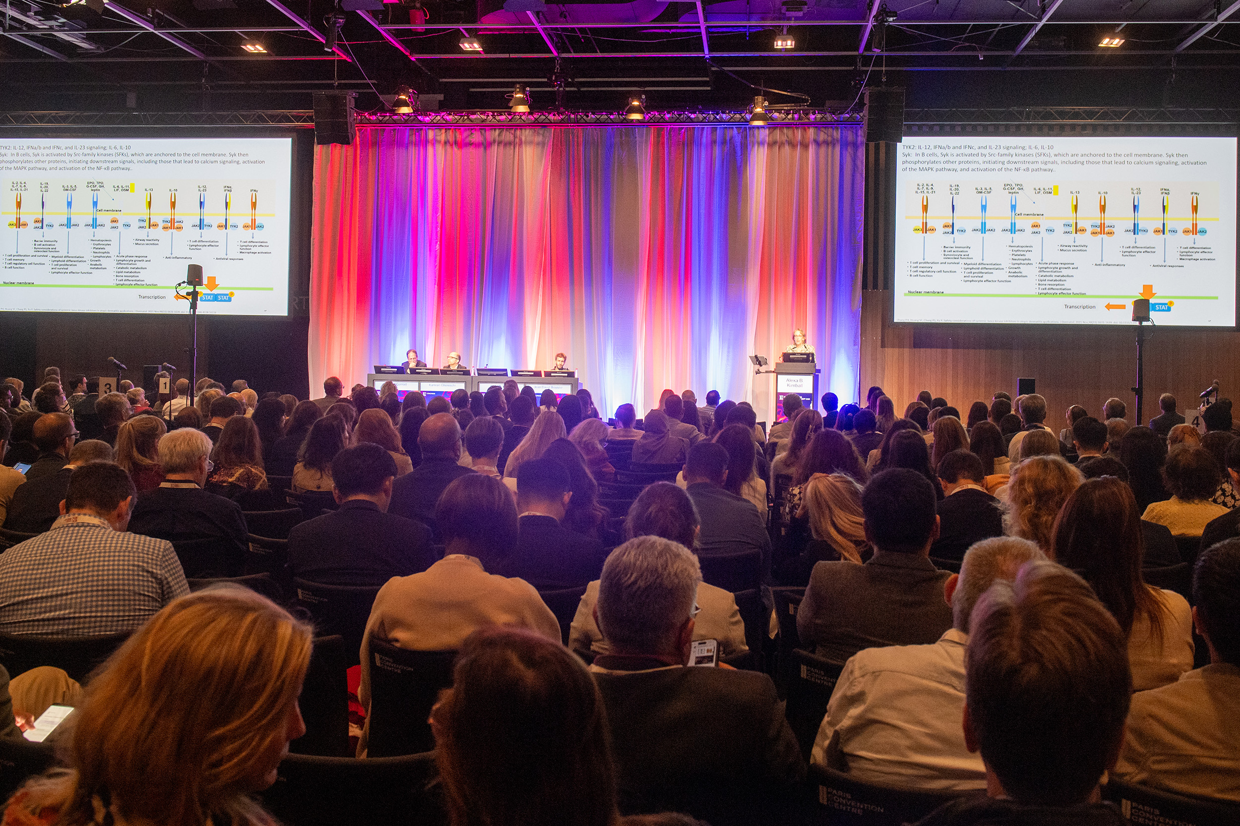
Prof. Alexa Kimball (Boston, US) presented on the role of JAK/TYK2 inhibitors in targeting IL-12, IL-23, type I interferons, and IL-6.
In dermatomyositis, phase 2/3 trials show early improvements in skin and muscle outcomes, although safety concerns such as cardiovascular and thrombotic risk remain. In hidradenitis suppurativa, pivotal trials demonstrated significant HiSCR50 responses by week 12, sustained through week 18, with a favourable safety profile.
Additional early data suggest potential benefits in prurigo nodularis, chronic prurigo, and lichen planopilaris.
JAK/TYK2 inhibitors hold promise across diverse skin diseases, but larger controlled studies are needed to confirm efficacy, define safety, and guide personalised treatment.
Prof. Denis Jullien
Thursday 18 September
Today's hot topics
Focus on hair disorders: Management
Androgenetic alopecia in children and adults
Dr. Luis Sanchez (Guadalajara, Mexico) opened the session by reviewing the current treatment options for androgenetic alopecia (AGA), characterised by a multifactorial pathogenesis and distinctive male and female pattern hair loss patterns.
AGA can present in children aged 6–9 years with a genetic susceptibility, and in post-menopausal women with andro-secreting tumours.
Management should be initiated as early as possible using combination therapies. Currently, only two approved pharmacological treatments are available: finasteride and minoxidil.
This condition can be distressing… patients may start self-induced behaviours such as trichotillomania.
Dr. Luis Sanchez
Anti-androgens are essential, although more data on their adverse effects (AEs) are needed, particularly regarding sexual dysfunction and neuropsychiatric disorders. Minoxidil has anti-inflammatory and antiandrogen benefits, with dose-dependent AEs (e.g. cardiovascular and hypertrichosis).
Emerging treatments including platelet-rich plasma therapy, new laser technologies (e.g. 675 nm), microneedling followed by exosome application, and hair transplantation offer new perspectives. PP405, a mitochondrial pyruvate carrier inhibitor, may be beneficial but more data are needed. Treatment adjustment is needed for some populations (e.g. children/adolescents, pregnant/lactating women, breast cancer or Transgender patients).
Dr. Sanchez highlighted the importance of a good patient-physician relationship to outline realistic, Individualised treatment outcomes, discuss different therapeutic alternatives and potential AEs.
Alopecia areata in children and adults
Dr. David Saceda Corralo, MD, PhD (Madrid, Spain) provided an overview of alopecia areata (AA), an established classical condition, to determine how AA can be better treated in the clinic.
AA is systemic autoimmune disease in which the immune system attacks hair follicles, resulting in nonscarring hair loss, affecting around 2% of the population. AA-related hair loss has many ‘faces’ or forms of clinical presentation. Severity assessment using the Severity of Alopecia Tool (SALT) score is fundamental to determine the amount of fair loss and individualise treatment.
Time is hair. As dermatologists we must know that the sooner you treat your patients, the better outcomes we will have.
David Saceda Corralo, MD, PhD
Mild, moderate and severe forms are treated differently, with severe AA typically requiring Janus kinase (JAK) inhibitor therapy. In addition, acute disease (rare; with AA in the last 6 months) can be treated with oral/systemic corticosteroids while chronic disease (most common) can be treated with JAK inhibitors.
Dr. Saceda Corralo noted that treatment options have changed the way physicians can treat their patients, and that results can take time. Some patients may be late responders and treatment should be continued, particularly in severe cases, to show a positive response. Combination treatment with baricitinib may also be useful for partial/non-responders or relapse. Red flags for optimal treatment outcomes include a SALT score >95 (total AA), an episode lasting >4 years and male gender.
However, JAK-inhibitors has shown sustained efficacy, and medical treatment should not be stopped unless there is a medical reason or the patient requests it.
Lichen planopilaris and frontal fibrosing alopecia
In his presentation, Prof. Jerry Shapiro (New York, US) focused on lymphocytic scarring alopecias.
Lichen planopilaris (LPP) is characterised by a gradual hair loss in adults, with itching (an early sign), pain, burning and shedding. Key trichoscopic features include inflamed skin, loss of follicular ostia, perifollicular scaling and pigment incontinence (blue-grey dots). Choice of LPP therapy is determined by the extent of the disease (</≥ 10%), and progression using an established treatment algorithm with standardised and additional treatment options. Rapidly-progressive forms are treated with oral prednisolone while combination therapy is recommended for less-rapid, intralesional LPP. Importantly, low-dose (anti-inflammatory) doxycycline twice-daily is as effective as the antimicrobial dose to improve AEs. Oral therapies (naltrexone and pioglitazone) and laser/light therapy are additional options.
I don’t recommend hair transplants (in these patients) …I’ve seen it start from a hair transplant…and to avoid eyebrow lifts or any facelift.
Prof. Jerry Shapiro
Frontal fibrosing alopecia (FFA) is distinguished by gradual hair loss of the outer canthus to the hairline in older adults, more commonly in women, with eyebrow involvement. Trichoscopic features include flattened hair shafts twisting around their own axis and a positive pull-test on edges of affected areas during active phase. Most patients can be treated and choice is based on treatment algorithm recommendations for slow or rapid progressive forms. Current therapies include topicals (e.g. tacrolimus, clobetasol, minoxidil, independently or in combination); intralesional triamcinolone acetonide, (although caution should be used to avoid central serous chorioretinopathy); with oral isotretinoin and topical JAK inhibitors to treat facial papules. Prof. Shapiro emphasised that some sunscreens and facial moisturisers can have a detrimental effect, and that hair transplants or brow lifts should be avoided.
Dissecting cellulitis and folliculitis decalvans
In the last session, Prof. Awatef Kelati (Casablanca, Morocco) outlined the challenges of treating chronic, inflammatory scalp conditions.
Dissecting cellulitis (DC) and folliculitis decalvans (FD) are both neutrophilic primary cicatricial alopecias with a chronic, relapsing course. Early identification of primary lesions can help to distinguish between the two diseases. In FD, the follicular pustule is typically as a result of bacterial colonisation, while follicular dilatation and keratin plugs in DC leads are part of an abnormal immune response. Trichoscopy correlates with pathophysiology and histopathology with FD located in the dermis and DC in the subcutis layers.
Folliculitis decalvans and dissecting cellulitis are more than just a scalp condition.
Prof. Awatef Kelati
DC provides a window into a systemic tendency for follicular occlusion and severe inflammation. Recognising the typical comorbidities (e.g. follicular involvement, acne, metabolic syndrome and arthritis) is essential for comprehensive diagnosis
Early diagnosis and effective treatment are key to improving the patient’s overall quality of life, with counselling to avoid scratching and/or further trauma to the skin). Similarly, early treatment of FD is important to prevent total destruction of hair follicles, leading to scarring alopecia.
Therapeutic options include topical treatment, intralesional corticosteroids, systemic antibiotics, and oral retinoids (i.e. isotretinoin). Additional therapies, including photodynamic therapy are interesting but more data are warranted. Prof. Kelati underlined that combination treatment leads to improvements in almost all cases and maintenance treatment is important to avoid relapse, particularly in young patients.
Key Takeaways:
- Treatment should be started as early as possible using combination therapy where indicated.
- JAK-inhibitors have shown sustained efficacy for chronic and severe AA
- Established treatment algorithms are available to select appropriate therapies for lymphocytic scarring alopecias.
- Maintenance treatment is important to avoid relapse.
Atopic dermatitis
New and Upcoming Treatments in AD
Prof. Emma Guttman-Yassky (New York, US) opened the session with a description of the shift from broad immunosuppressants to targeted therapies for type 2 inflammation in atopic dermatitis. She noted durable benefits from dual IL-4/IL-13 blockade, with IL-13–specific inhibition showing slightly lower responses. Targeting IL-31 offers rapid relief of itch and sleep disturbance, while OX40–OX40L modulation may reprogramme T-cell activity for longer-term remission.
She also noted that oral small molecules blocking multiple pathways provide convenience and efficacy, though safety monitoring remains critical.Phase 3 data in giant cell arteritis demonstrated reduced flare rates, while safety analyses covering >27,000 patient-years revealed no new signals.
Prof. Ghoreschi also pointed to biomarker studies linking immune pathway modulation with clinical regrowth in alopecia and four-year psoriasis extension data confirming durable PASI responses.
Role of environment and allergens in AD flares
Prof. Matthias Schmuth (Innsbruck, Austria) highlighted that atopic dermatitis flares are not solely driven by IL-4/IL-13 signalling but also by a basophil-mediated neuroimmune circuit, with allergen-stimulated basophils releasing leukotriene C4 to activate sensory nerves and trigger itch. He underscored the critical role of environmental factors in barrier dysfunction and flare risk, with heat, low humidity, pollutants, tobacco smoke, water hardness, and detergents all impairing skin integrity and amplifying inflammation.
Modifying environmental exposures, ensuring regular emollient use, and strengthening patient education are key strategies for prevention, as reflected in the 2025 EuroDerm guidelines.
Prof. Matthias Schmuth
Super-responders and Disease Modification in AD
Prof. Eric Simpson (Oregon, US) discussed the concept of super-responders in atopic dermatitis, defined as patients achieving EASI ≤7, PP-NRS ≤4, DLQI ≤5, and POEM ≤7 at week 16 and maintaining this through one year. He noted that such patients may be candidates for dose reduction, extended dosing intervals, or even treatment withdrawal. Early intervention, particularly in children, may not only control skin disease but also reduce or prevent comorbidities such as asthma, allergies, growth impairment, and mental health issues.
Prof. Simpson highlighted the emerging idea of disease modification, in which treatment durably alters the natural course of AD, with patients maintaining control even off therapy. He concluded that early targeted intervention may support normal growth and lower the risk of the atopic march, although standardised remission criteria and predictive biomarkers are still needed.
Difficult to treat and treatment-resistant AD
Prof. Thomas Bieber (Davos, Switzerland) addressed the management of therapy-resistant and difficult-to-treat atopic dermatitis, emphasising the importance of distinguishing true resistance from cases that only appear resistant due to misdiagnosis, poor adherence, psychological triggers, infections, or environmental factors. He urged clinicians to reconsider diagnoses, investigate possible “chameleons,” and use tools such as biopsy, patch testing, and adherence checks.
Prof. Bieber stressed that management requires a personalised, patient-centred approach. Biologics and JAK inhibitors can play an important role, particularly in patients with phenotype switch (“flip-flop” AD) or complex comorbidities, but shared decision-making, education, and close monitoring remain essential for optimal outcomes.
There is no one-size-fits-all for complex diseases.
Prof. Thomas Bieber
Key Takeaways:
- Targeted therapies against type 2 inflammation, novel pathways such as IL-31 and OX40–OX40L, and oral small molecules are reshaping AD management.
- Beyond drugs, addressing environmental triggers, ensuring adherence, and tailoring therapy to comorbidities or phenotype switch remain central to AD management.
Bullous diseases
Dermatitis herpetiformis
Prof. Agnieszka Żebrowska (Białystok, Poland) described dermatitis herpetiformis as a cutaneous manifestation of coeliac disease and a clear example of the gut–skin axis.
Diagnosis rests on direct immunofluorescence and anti-TG3 antibodies, supported by serology, while atypical presentations may resemble urticaria, prurigo, or palmo-plantar lesions. Management is centred on strict adherence to a gluten-free diet, with additional pharmacological options available for symptom control and biologic therapies reserved for refractory cases. New frontiers in dermatitis herpetiformis include biologics, biomarkers, probiotics, and AI-driven personalised approaches.
Bullae in Other Skin Diseases
Prof. Emiliano Antiga (Florence, Italy) reviewed blistering diseases beyond classic autoimmune causes, classifying them by topographic and etiopathologic features.
He highlighted genetic disorders, infectious origins, and metabolic, nutritional, and photosensitive conditions, as well as physical triggers such as burns, insect bites, and friction. Drug reactions, including Stevens-Johnson syndrome and toxic epidermal necrolysis, and inflammatory diseases such as erythema multiforme, bullous Sweet syndrome, and eosinophilic dermatosis of haematologic malignancies, were also discussed.
Bullae are not always autoimmune in origin. Clinicians should be cautious with atypical cases, and when in doubt, confirm with direct immunofluorescence.
Prof. Emiliano Antiga
Epidermolysis bullosa acquisita (EBA)
Prof. Barbara Horváth (Groningen, Netherlands) described EBA as a rare, chronic autoimmune blistering disease caused by autoantibodies to type VII collagen. It presents in two main forms: mechanobullous, with trauma-induced scarring blisters, and inflammatory, resembling bullous pemphigoid; both may involve mucosa.
Diagnosis relies on direct and indirect immunofluorescence and serology, while associations with autoimmune and haematologic comorbidities add to its complexity. Management follows a stepwise approach from anti-inflammatory and immunomodulatory therapy to immunosuppression and biologics in refractory cases. Despite its chronic course, Prof. Horváth explained that mortality is generally favourable and emerging targeted therapies may improve outcomes.
Special focus: Children
Dr. Carmen Sălăvăstru (Bucharest, Romania) discussed vesiculobullous disease in children, highlighting its broad differential and the need for careful history, examination, and labs to separate benign from life-threatening cases. Neonatal autoimmune blistering (e.g., pemphigus, bullous pemphigoid) typically resolves with declining maternal antibodies, except chronic bullous disease of childhood, which may require stopping breastfeeding and systemic therapy.
In older children, juvenile bullous pemphigoid often affects the extremities and may need stepwise treatment from corticosteroids to biologics. Genetic disorders like epidermolysis bullosa require long-term care focusing on inflammation, pruritus, and wound management. She concluded with a practical diagnostic algorithm for decision-making and triage.
Vesiculobullous lesions in children can signal a wide range of genetic, autoimmune, infectious, metabolic, or traumatic diseases. A structured diagnostic approach is essential to distinguish benign conditions from life-threatening blistering disorders.
Dr. Carmen Sălăvăstru
Key Takeaways:
- Bullous disorders extend well beyond classic autoimmune causes, encompassing genetic, infectious, metabolic, nutritional, drug-related, and traumatic conditions.
- Management strategies are evolving from strict dietary and symptomatic control to stepwise immunomodulation, with biologics and emerging tools such as biomarkers, microbiome modulation, and AI offering new opportunities for refractory or complex cases.
Nail disorders
Nail psoriasis
Prof. Matilde Iorizzo (Lugano, Switzerland) highlighted nail psoriasis as a common but underestimated manifestation, affecting up to 60% of patients and sometimes predicting more severe disease. She reviewed its clinical signs, Th17-driven inflammation, and links to psoriatic arthritis, stressing the need to distinguish it from onychomycosis using nail clippings, histology, and ultrasound.
Prof. Iorizzo explained how management is challenging due to slow nail growth, but new topical compounds, IL-17 and IL-23 inhibitors, and oral IL-23 agents are under investigation. She concluded that early recognition, precise diagnosis, and persistence are essential to improving long-term outcomes.
Drug-induced nail changes
Dr. Tomer Goldsmith (Tel Aviv, Israel) described drug-induced nail changes as sentinel signs of systemic drug reactions, which may mimic systemic disease, infections, or neoplasms.
He reviewed their varied presentations, including pigmentary, structural, vascular, and inflammatory or periungual changes, and explained that underlying mechanisms range from direct toxicity and pigment deposition to immune-mediated injury, vascular occlusion, and phototoxicity. Assessing timing and dose-dependency is critical for causality. Management is largely supportive, focusing on protective measures, nail care, and monitoring for secondary infection, with drug withdrawal reserved for severe cases.
Dr. Goldsmith concluded that recognising drug-related nail changes is essential to prevent misdiagnosis
Drug reactions in the nails, much like in the skin, can mimic almost any disease and may even precede other adverse manifestations, making early recognition vital.
Dr. Tomer Goldsmith
Lichen planus
Prof. Bertrand Richert (Brussels, Belgium) opened his presentation with the message that nail lichen planus (NLP) is a “true nail emergency,” carrying the risk of permanent scarring and functional loss if not treated early. Clinical signs reflect matrix and nail bed involvement, and diagnosis requires biopsy with exclusion of mimics.
Prof. Richert noted that topical therapy is generally ineffective, making aggressive treatment essential: intralesional therapy can help in limited disease, systemic therapy may achieve control (although relapse is common), and newer targeted approaches are emerging for refractory cases.
Prof. Richert concluded that early recognition, biopsy, and decisive management are critical to improving outcomes.
Special focus: Children
Dr. Lourdes Navarro Campoamor (Madrid, Spain) presented on nail alterations in children, noting that while many, such as periungual hyperkeratosis in newborns, are benign, careful assessment is essential to avoid misdiagnosis.
Retronychia, now seen as a dynamic process often linked to malalignment, benefits from early recognition to prevent damage. Onychomycosis remains underdiagnosed, underscoring the need for family-wide, risk-based management.
Paediatric melanonychia is usually benign and best handled with shared decision-making, dermoscopy, and monitoring rather than biopsy.
Nail changes also feature in genetic disorders like ichthyosis, reflecting genotype–phenotype links. Dr. Navarro Campoamor concluded by stressing the importance of vigilance, early detection, and patient-centred care.
Fungal infections in children remain underestimated and more robust data are needed.
Dr. Lourdes Navarro Campoamor
Key Takeaways:
- From nail psoriasis and lichen planus to drug-induced and paediatric nail changes, timely diagnosis is essential to prevent irreversible damage, avoid misdiagnosis, and ensure appropriate management.
- Nail disease care remains challenging, with many cases requiring long-term monitoring and patient-centred strategies. While supportive and conventional treatments are still mainstays, emerging therapies and targeted approaches are expanding future options and improving prospects for better outcomes.
Personalised approaches for clinical practice
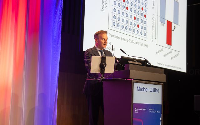
Molecular profiling
Prof. Michel Gilliet (University Hospital Lausanne, Switzerland) opened the session with a timeline of the molecular revolution in dermatology. He explained how mapping dominant immune pathways (Th1, Th2, Th17, IFN-I) improves diagnosis, unmasks misdiagnosis, and guides targeted therapy in diseases such as psoriasis, atopic dermatitis, lupus, lichen planus, and palmoplantar disorders.
He also showed clinical examples of how immune signatures can distinguish between similar conditions, predict treatment responses, and explain resistance or paradoxical reactions through immune shifts.
Molecular profiling of inflammatory skin diseases enables accurate diagnosis in challenging cases, enhances therapeutic efficacy, clarifies causes of treatment failure, and informs therapy selection for non-responding patients.
Prof. Michel Gilliet
Personalised drug dosing
Dr. Jo Lambert (Ghent University, Belgium) presented on the need for personalised drug dosing in dermatology.
She explained that biologics have transformed treatment for chronic inflammatory dermatoses, but “one-size-fits-all” dosing fails to reflect patient heterogeneity. Therapeutic drug monitoring can help, yet trough-based approaches are limited and impractical in self-administered settings. Population PK/PD models with Bayesian forecasting provide a more precise solution by combining prior data with real-time patient measurements to guide dosing.
Dr. Lambert concluded that model-informed precision dosing could improve outcomes, reduce costs, and better tailor therapy, although reimbursement and implementation challenges remain.
Team medicine approach
Dr. Kåre Steinar Tviet (Bergen, Norway) drew on lessons from team building in the aviation industry to emphasise the importance of a “team medicine” approach in dermatology.
He highlighted strong evidence linking teamwork culture to reduced burnout, fewer adverse events, and improved patient safety. He emphasised that effective teams depend on clear roles, mutual trust, psychological safety, and strong leadership that promotes open communication and collaboration.
Through examples such as transplantation, vulva clinics, Mohs surgery, rare disease networks, and emergency responses for severe skin reactions, he illustrated how this approach improves outcomes.
Shared decision making
Dr. Hélène Aubert Wastiaux (Nantes University Hospital, France) highlighted the growing role of shared decision-making (SDM) in dermatology, particularly in conditions such as psoriasis and atopic dermatitis where systemic therapies often show similar efficacy.
She described SDM as a structured process in which patients and physicians exchange information, deliberate on options, and agree on a plan that integrates medical expertise with patient values. It reflects the broader shift toward patient empowerment, wider access to medical knowledge, and personalised care. Barriers to SDM include limited time, lack of continuity of care, and varying patient engagement, while facilitators include motivated clinicians, use of decision aids, and evidence that SDM enhances adherence, satisfaction, and outcomes.
Shared decision-making not only reduces decisional regret but also strengthens the therapeutic alliance, benefiting both patients and physicians.
Dr. Hélène Aubert Wastiaux
Key Takeaways:
- Molecular profiling and model-informed drug dosing are moving the field beyond “one-size-fits-all” treatment, enabling more accurate diagnosis, tailored therapy selection, improved efficacy, and cost efficiency.
- Strong teamwork cultures and structured shared decision-making both enhance safety, reduce burnout, strengthen the therapeutic alliance, and ensure care is aligned with patient values.
Hot topics in dermatology selected by the EADV President
How to reach unity in dermatology
Prof. Matthias Schmuth (Innsbruck, Austria) highlighted that atopic dermatitis flares are not solely driven by IL-4/IL-13 signalling but also by a basophil-mediated neuroimmune circuit, with allergen-stimulated basophils releasing leukotriene C4 to activate sensory nerves and trigger itch. He underscored the critical role of environmental factors in barrier dysfunction and flare risk, with heat, low humidity, pollutants, tobacco smoke, water hardness, and detergents all impairing skin integrity and amplifying inflammation.
Unity in dermatology is not about erasing differences – it is about harnessing them to heal, innovate, and serve.
Prof. Seemal Desai
Car T-cell therapy in autoimmune diseases
Dr. Marc Scherlinger (Strasbourg, France) reviewed the role of CAR-T cells in systemic autoimmune diseases.
He explained that current B-cell–directed therapies often spare long-lived plasma cells, whereas anti-CD19 CAR-T cells achieve deeper depletion and a partial repertoire reset. Dual CD19/BCMA targeting may also eliminate plasma cells but risks profound antibody loss. He highlighted that future directions, such as off-the-shelf CAR-T therapies, antigen-specific CARs like DSG3-CAART for pemphigus vulgaris, and T-cell engagers, are potentially simpler alternatives.
Dr. Scherlinger concluded that although costly and complex, CAR-based therapies hold strong potential for long-term remission in refractory autoimmune disease.
From tolerance to therapy: Harnessing inducible regulatory T-cells for precision control of autoimmune disease
Dr. Masayuki Amagai (Tokyo, Japan) presented new research on induced regulatory T cells (iTregs) for pemphigus vulgaris (PV), a blistering disease driven by autoantibodies against desmoglein-3 (Dsg3).
He explained that Dsg3-specific CD4⁺ T cells orchestrate autoantibody production when tolerance mechanisms fail. In mouse models, antigen-specific iTregs suppressed pathogenic T-cell responses, limited B-cell activation, and reduced anti-Dsg3 antibodies. A stabilised form (S/F-iTregs), engineered to maintain Foxp3, showed enhanced regulatory activity, antigen specificity, and significant disease suppression.
Dr. Amagai concluded that these advances pave the way for first-in-human studies and mark a shift from broad immunosuppression to precise, antigen-targeted therapy in autoimmune skin disease.
In 1991, during my fellowship at John Stanley’s lab at the NIH, I was fortunate to isolate the cDNA for the pemphigus vulgaris antigen, now known as desmoglein 3. This discovery helped establish that the pathophysiology of pemphigus lies in IgG autoantibodies disrupting keratinocyte adhesion, leading to blister formation.
Dr. Masayuki Amagai
Key Takeaways:
- There is a call for unity in dermatology – embracing diversity across cultures and specialties to strengthen advocacy, education, and patient care worldwide.
- Although costly and complex, CAR-T therapies can achieve deeper B-cell depletion and offer long-term remission for refractory autoimmune diseases.
- Antigen-specific iTregs, especially stabilised forms maintaining Foxp3, can suppress pathogenic immunity in pemphigus models and pave the way for targeted therapies
Dermoscopy for melanoma and melanocytic naevi
Indicators of melanoma in pigmented lesions
Prof. Giuseppe Argenziano (Naples, Italy) started the session with an engaging review of melanoma criteria in dermoscopy. Although seven main criteria have been recognised, additional criteria have been added over the last 30 years. He asked the participants how many were necessary before noting that in his opinion, one is enough to perform a biopsy.
The seven main criteria include: an atypical network, atypical vessels, blue-white veil, regression structures, irregular streaks, or reticular depigmentation in the form of dots/globules or blotches. The most important and most common is an atypical network, when more than one type is seen in the same lesion: however, there may be varying degrees with some between typical-atypical lesions. The other criteria listed are useful to provide additional clues.
He emphasised that regression (not palpable) is often seen in early melanoma, and that a blue colour is not specific for melanoma and other areas and combinations (the blue/black rule) are more important to suggest nodular melanoma.
In addition, criteria based on special locations such as melanoma in situ (network and regression) and white lines observed in Spitz neavus can be useful to differentiate dermal naevi versus melanoma.
One criterion is enough to say convincingly that this is a melanoma.
Prof. Giuseppe Argenziano
Spitzoid lesions
Prof. Bengü Nisa Akay (Ankara, Turkey) gave her insights into a challenging area for dermatologists.
Spitzoid lesions are a controversial group of tumours in terms of their clinical and dermatoscopic features, and management strategies. They represent 1–2% of all tumours and evolve spontaneously over time. It can be difficult to determine how many are true melanomas without a patient history.
The clinical differential should be made cautiously, especially in children.
Prof. Bengü Nisa Akay
Next-generation sequencing (NGS) is useful to differentiate between different lesion types, which are defined as: Spitz naevus (SN) atypical Spitz tumour (AST)/Spitz melanocytoma) or a Spitz melanoma (SM). SM is reported more frequently in males, and all types are more common in white patients.
Key indicators of melanoma, determined using immunohistochemistry, include: high Ki-67 index in the dermisloss of p16 expression, and strong preferentially-expressed antigen in melanoma positivity. NGS and fluorescent in-situ hybridisation are required for challenging cases.
Prof. Akay recommended that asymmetric or post-pubertal Spitz-like lesions should always be excised, irrespective of morphology, with symmetry and patient age as primary decision-makers for biopsy or monitoring. Sentinel lymph node biopsy is not usually indicated in Spitz nevus or AST unless melanoma is strongly suspected.
She concluded by highlighting that Spitz melanomas with kinase fusions tend to behave less aggressively than Spitzoid melanomas driven by the BRAF or NRAS mutations.
Lentigo maligna
Dr. Jilliana Monnier (Marseille, France) shared her experience on Lentigo maligna, (LM), which is inherently difficult to diagnose and treat in the clinic.
LM is characterised by a slow-growing, malignant and invasive lesion on sun-exposed skin, typically on the nose, which has the ability to metastasise.
A small lesional size may make LM difficult to distinguish from other pigmented benign lesions (e.g. Seborrhoeic keratosis and Solar lentigo). An asymmetric pigmented follicular opening (illustrated on Dermoscopedia.org) and angulated lines are more relevant as indicators for LM, which can be examined using high-magnification dermoscopy to help make a diagnosis.
Lesions on the face are really difficult because (patients) ask for treatment, lasers, creams…and we don’t want to miss Lentigo maligna.
Dr. Jilliana Monnier
In recent years, high-magnification imaging tools with integrated artificial intelligence systems have been developed to further aid diagnostic accuracy and excision and may enable automated melanoma screening in the future. Newer multimodal imaging techniques (e.g. reflectance confocal microscopy and line-field confocal optical coherence tomography) are already in use enabling both diagnosis and margin delimitation (which is often difficult to treat), and may help to improve clinical management.
Physicians need to be cautious of infiltrative, invasive LM where punch biopsies should not be used as they would give an incorrect diagnosis.
Pigmented lesions of the oral and genital mucosa
Prof. Monika Arenbergerova (Prague, Czech Republic) began by asking attendees if they regularly examined patients with pigmented lesions on the body, face, acral area, under the nails, or genitals before noting that the latter is often left unchecked.
Diagnosis and treatment of oral and genital lesions represent several challenges as currently only a few studies are available, and there is often significant patient embarrassment associated with dermoscopic examination of the genital area. In addition, there is a lack of specific dermoscopic equipment to enable visualisation of intraoral lesions.
This is a challenging topic…we need new algorithms.
Prof. Monika Arenbergerova
Pigmented mucosal lesions are rare, and are typically diagnosed by assessing the anatomical site, pattern, colour and type of lesion, which can be benign or malignant, constitutive or acquired. Melanoma of the lips or oral cavity is the least common, while pigmented lesions on the penile and vulval areas follow distinctive patterns, most typically a ring-like pattern. There are eight different dermoscopy patterns for vulval lesions. However, while traditional algorithms for dermoscopy are available, they aren’t effective for clinical practice.
Prof. Arenbergerova underlined that biopsy or excision should be always performed when a suspected lesion meets one of the following three criteria:
a structureless area with a blue, gray or white colour; where the predominant colour is blue, white or gray only; or a new lesion in patients aged 40 years or more.
Importantly, different immunotherapy combinations can improve overall 5-year survival rates in patients with malignant mucosal melanomas.
Key Takeaways:
- An atypical network is indicative of a suspected melanoma in pigmented lesions.
- Next-generation sequencing is integral to differentiating Spitzoid lesion types.
- High-magnification imaging with integrated artificial intelligence systems will further aid diagnostic accuracy and excision of LM.
- Combination therapy may improve prognosis in pigmented oral/genital lesions.
Acne
Acne guidelines 2025: Critical assessment
Dr. Neal Bhatia, M.D (San Diego, US) set the scene for this session by appraising the current guidelines for acne with his own observations.
Key highlights include the reclassification of Propionibacterium acnes (P. acnes) to Cutibacterium acnes (C. acnes), and using new pathogenic models to understand where novel therapeutic strategies should be directed to reduce scarring. Diet can affect inflammatory pathways in acne and targeting the Th17 pathway may help to reduce inflammation.
Make sure you take good pictures before and after so you know where you’re going to end up.
Dr. Neal Bhatia, M.D
Key takeaways from the guidelines included the ‘Triple Threat’ combination approach (using clindamycin phosphate, benzoyl peroxide, and adapalene); assessing hormonal acne as acne; and use of isotretinoin is integral to effective treatment (physicians should not be deterred by social media). Although not recommended, minocycline is an effective anti-inflammatory topical agent when used with adjuncts for a limited time.
Dermatologists should optimise topical treatments, particularly in severe disease when combined with systemic treatment. Newer therapeutic formulations and innovations (e.g. cannabinoids, zinc, red light photo dynamic therapy and 1726 laser energy devices) can be useful and should be supported where efficacy and tolerability are greater than generic treatment.
The foundation and key priority of management is to improve erythema for the patient.
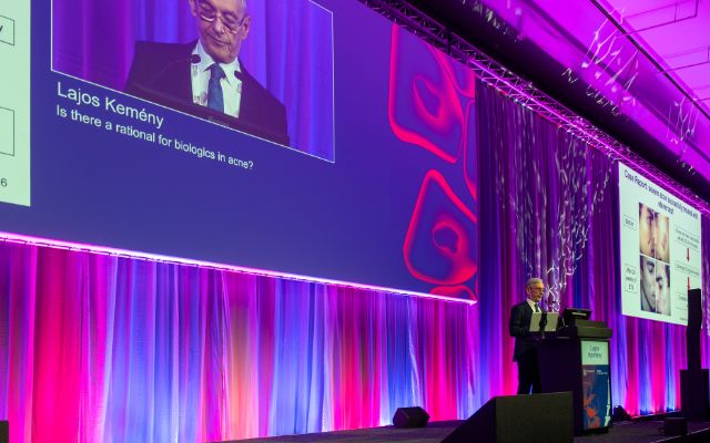
Is there a rationale for biologics in acne?
Prof. Dr. Lajos Kemény (Szeged, Hungary) then outlined the potential for using biologics in acne.
No biologics are currently approved for acne; to date only three clinical trials have been conducted which failed to demonsrate differences versus placebo. This may have been a result of these therpaies being started too late, or there is a need to target other cytokine pathways, which may lead to new reseearch in the future.
We may have some new data (on biologics) in the future, but we are not there yet.
Prof. Dr. Lajos Kemény
Although postive results have been reported in several case reports, where antibiodies were shown to be effectivive in block cytoklines, caution should be used in interpretating these results. For therapy-resistant acne, it is important to consider a ’conglobate type’ and if facial localisation of hidradenitis suppurativa is pressent, not acne.
For severe, therapy-resistant acne with or without ’acne syndrome’, dermatologists can consider tumour necrosis factor α, interleukin (IL)-17 or IL-23 inhibitors. Use of other bacterial strains to modulate the skin microbiome, neutralising antibodies against C. acnes virulence factors and phage therapy to kill C. acnes may be useful to improve acne.
Artificial Intelligence to score acne severity
Dr. Vincenzo Bettoli MD (Ferrara, Italy) discussed the importance of artificial intelligence (AI) in the acne setting.
It is critical to first assess acne severity correctly before deciding on the most appropriate treatment, to assess treatment results, with good reproducibility. Key challenges include the different lesional types (acute phase and sequellae), and that lesions may be uneven and in continuous evolution with spontaneous fluctuation in different sites. Three-dimensional evaluation is critical.
AI is far from being useful is this specific aspect (of treatment) so far.
Dr. Vincenzo Bettoli MD
Although a number of grading systems have been proposed for acne, the ‘perfect’ grading system has not yet been developed. In the advent of newer technologies, initial computerised AI approaches have been developed to identify acne lesions. However, limitations including data imbalances and lack of patient diversity have impeded their accuracy, generalisability and reproducibility. External validation by dermatologists is still needed.
Prospective studies in real-world settings are required to validate the effectiveness of the algorithms and the AI training of these systems in dermatology. Dr Bettoli emphasised that acne severity, medical history and discomfort – alongside patient preference – are the cornerstones of forming personalised and effective therapeutic decisions. He cautioned that further developments are needed for AI to be useful in this setting.
Management of post-acne hyperpigmentation
In the last presentation, Prof. Dr. Jose Luis Lopez-Estebaranz (Madrid, Spain) reviewed how hyperpigmentation can be managed.
Although there are few reports in the literature acne vulgaris affects many individuals and may lead to post-inflammatory erythema and post-inflammatory hyperpigmentation, particularly in females with darker skin (Fitzpatrick types III–VI). Risk factors include severe acne, excessive sun exposure, squeezing/scratching lesions and facial site. The impact on quality of life underscores the need for effective treatment.
Add moisturiser and a high (broad-spectrum sun) protection…as you know, visible light is what impacts more to produce hyperpigmentation.
Prof. Dr. Jose Luis Lopez-Estebaranz
Acne should be treated effectively and promptly without causing irritation or damage to the skin barrier. Recommended therapeutic options are isotretinoin with photoprotection and trifarotene, and use of triple combinations to reduce inflammatory lesions. Additional agents include azelaic acid, tranexmic acid and hydroquinone (but to use with care to avoid hypopigmentation) and cysteamine, a newly-emerging agent.
Additional therapies such as chemical peels, energy-based devices to improve hyperpigmentation (e.g. non-ablative laser and antioxidants, noting that Ruby lasers should not be used); and radiofrequency microneedling can be used with good results. Good skincare and topical treatments, ensuring hydration before and after treatment, are also fundamental.
Key Takeaways:
- Propionibacterium acnes has been reclassified to Cutibacterium acnes in the most recent guidelines.
- No biologic therapies have been approved for acne; targeting other pathways may be useful.
- AI is not yet useful: severity, medical history, discomfort and patient preference are crucial.
- Post-acne hyperpigmentation treatment should be initiated promptly and effectively, with optimal hydration.
Chronic prurigo
Understanding chronic prurigo
Dr. Manuel Pereira (Berlin, Germany) opened the session with a comprehensive overview of chronic prurigo (CPG), a debilitating condition characterised by persistent itch lasting at least six weeks, repeated scratching, and the development of pruriginous lesions.
He highlighted the complex interplay of immunological and neuronal mechanisms underlying the disease, with cutaneous studies revealing reduced intraepidermal nerve fibre density and enhanced neuronal branching that amplify itch perception.
Dr. Pereira emphasised the heavy burden CPG places on patients, with visible lesions, scarring, and profound impacts on quality of life.
Innovative therapies targeting the neuro-immune axis show promise for the treatment of chronic prurigo.
Dr. Manuel Pereira
Diagnostic workup
Prof. Jacek Szepietowsky (Wroclaw, Poland) focused on the diagnostic workup of CPG, stressing that while the condition is frequently encountered in daily practice, accurate diagnosis remains challenging. He outlined the need for a structured approach that considers medical history, comorbidities, physical and laboratory examinations, and the psychosocial burden of disease.
Tools such as itch intensity scales, questionnaires, and scratch activity measures can support evaluation, but each has limitations.
Crucially, diagnosis requires differentiating CPG from other causes of chronic pruritus.
Diagnosis and assessment of chronic prurigo is a clinical challenge, requiring a holistic approach and experienced clinical judgement.
Prof. Jacek Szepietowsky
Emerging biologics
Prof. Sonja Ständer (University Hospital of Münster, Germany) discussed advances in biologic therapies for CPG.
She explained that the disease involves barrier dysfunction, neuroimmune interactions, type 2 inflammation, and fibrosis. Clinical trials show that biologics targeting interleukin pathways can provide rapid itch relief, promote lesion healing, and significantly improve quality of life, with sustained benefits observed in long-term studies.
Early-phase data also suggest promise for therapies directed at additional immune and mast cell pathways, although long-term safety and treatment optimisation remain key priorities.
Emerging oral therapies
Dr. Gil Yosipovitch (Miami, Florida) spoke about the central role of neural and immunologic mediators such as IL-4, IL-13, and IL-31 in driving itch and lesion development in prurigo nodularis (PN), making them key therapeutic targets.
He highlighted that JAK inhibitors have demonstrated rapid and clinically meaningful improvements in both itch and lesions, often within weeks and in some cases faster than biologics.
Extension studies further support the durability of these responses. However, Dr. Yosipovitch emphasised that safety considerations remain important, particularly in older PN patients with comorbidities.
JAK–STAT inhibitors hold promise for PN, especially in refractory cases, but long-term safety data are limited, optimal dosing is unclear, and head-to-head biologic trials are needed.
Dr. Gil Yosipovitch
Key Takeaways:
- CPG is a complex neuro-immune disease with a heavy patient burden that underscores the need for accurate diagnosis and holistic management.
- New therapeutic avenues are emerging, with biologics and oral therapies targeting the neuro-immune axis showing rapid and durable improvements in itch and lesions.
Dermoscopy for melanoma and melanocytic naevi
Indicators of melanoma in pigmented lesions
Prof. Giuseppe Argenziano (Naples, Italy) started the session with an engaging review of melanoma criteria in dermoscopy. Although seven main criteria have been recognised, additional criteria have been added over the last 30 years. He asked the participants how many were necessary before noting that in his opinion, one is enough to perform a biopsy.
The seven main criteria include: an atypical network, atypical vessels, blue-white veil, regression structures, irregular streaks, or reticular depigmentation in the form of dots/globules or blotches. The most important and most common is an atypical network, when more than one type is seen in the same lesion: however, there may be varying degrees with some between typical-atypical lesions. The other criteria listed are useful to provide additional clues.
He emphasised that regression (not palpable) is often seen in early melanoma, and that a blue colour is not specific for melanoma and other areas and combinations (the blue/black rule) are more important to suggest nodular melanoma.
In addition, criteria based on special locations such as melanoma in situ (network and regression) and white lines observed in Spitz neavus can be useful to differentiate dermal naevi versus melanoma.
One criterion is enough to say convincingly that this is a melanoma.
Prof. Giuseppe Argenziano
Spitzoid lesions
Prof. Bengü Nisa Akay (Ankara, Turkey) gave her insights into a challenging area for dermatologists.
Spitzoid lesions are a controversial group of tumours in terms of their clinical and dermatoscopic features, and management strategies. They represent 1–2% of all tumours and evolve spontaneously over time. It can be difficult to determine how many are true melanomas without a patient history.
The clinical differential should be made cautiously, especially in children
Prof. Bengü Nisa Akay
Next-generation sequencing (NGS) is useful to differentiate between different lesion types, which are defined as: Spitz naevus (SN) atypical Spitz tumour (AST)/Spitz melanocytoma) or a Spitz melanoma (SM). SM is reported more frequently in males, and all types are more common in white patients.
Key indicators of melanoma, determined using immunohistochemistry, include: high Ki-67 index in the dermisloss of p16 expression, and strong preferentially-expressed antigen in melanoma positivity. NGS and fluorescent in-situ hybridisation are required for challenging cases.
Prof. Akay recommended that asymmetric or post-pubertal Spitz-like lesions should always be excised, irrespective of morphology, with symmetry and patient age as primary decision-makers for biopsy or monitoring. Sentinel lymph node biopsy is not usually indicated in Spitz nevus or AST unless melanoma is strongly suspected.
She concluded by highlighting that Spitz melanomas with kinase fusions tend to behave less aggressively than Spitzoid melanomas driven by the BRAF or NRAS mutations.
Lentigo maligna
Dr. Jilliana Monnier (Marseille, France) shared her experience on Lentigo maligna, (LM), which is inherently difficult to diagnose and treat in the clinic.
LM is characterised by a slow-growing, malignant and invasive lesion on sun-exposed skin, typically on the nose, which has the ability to metastasise.
A small lesional size may make LM difficult to distinguish from other pigmented benign lesions (e.g. Seborrhoeic keratosis and Solar lentigo). An asymmetric pigmented follicular opening (illustrated on Dermoscopedia.org) and angulated lines are more relevant as indicators for LM, which can be examined using high-magnification dermoscopy to help make a diagnosis.
Lesions on the face are really difficult because (patients) ask for treatment, lasers, creams…and we don’t want to miss Lentigo maligna.
Dr. Jilliana Monnier
In recent years, high-magnification imaging tools with integrated artificial intelligence systems have been developed to further aid diagnostic accuracy and excision and may enable automated melanoma screening in the future. Newer multimodal imaging techniques (e.g. reflectance confocal microscopy and line-field confocal optical coherence tomography) are already in use enabling both diagnosis and margin delimitation (which is often difficult to treat), and may help to improve clinical management.
Physicians need to be cautious of infiltrative, invasive LM where punch biopsies should not be used as they would give an incorrect diagnosis.
Pigmented lesions of the oral and genital mucosa
Prof. Monika Arenbergerova (Prague, Czech Republic) began by asking attendees if they regularly examined patients with pigmented lesions on the body, face, acral area, under the nails, or genitals before noting that the latter is often left unchecked.
Diagnosis and treatment of oral and genital lesions represent several challenges as currently only a few studies are available, and there is often significant patient embarrassment associated with dermoscopic examination of the genital area. In addition, there is a lack of specific dermoscopic equipment to enable visualisation of intraoral lesions.
This is a challenging topic…we need new algorithms.
Prof. Monika Arenbergerova
Pigmented mucosal lesions are rare, and are typically diagnosed by assessing the anatomical site, pattern, colour and type of lesion, which can be benign or malignant, constitutive or acquired. Melanoma of the lips or oral cavity is the least common, while pigmented lesions on the penile and vulval areas follow distinctive patterns, most typically a ring-like pattern. There are eight different dermoscopy patterns for vulval lesions. However, while traditional algorithms for dermoscopy are available, they aren’t effective for clinical practice.
Prof. Arenbergerova underlined that biopsy or excision should be always performed when a suspected lesion meets one of the following three criteria:
a structureless area with a blue, gray or white colour; where the predominant colour is blue, white or gray only; or a new lesion in patients aged 40 years or more.
Importantly, different immunotherapy combinations can improve overall 5-year survival rates in patients with malignant mucosal melanomas.
Key Takeaways:
- An atypical network is indicative of a suspected melanoma in pigmented lesions.
- Next-generation sequencing is integral to differentiating Spitzoid lesion types.
- High-magnification imaging with integrated artificial intelligence systems will further aid diagnostic accuracy and excision of LM.
- Combination therapy may improve prognosis in pigmented oral/genital lesions.