
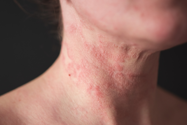
Atopic dermatitis in adults
UPDATES
- Friday, 02 October
- 10:15 - 11:45 CEST
- Hall A
Presentation details:

Late-onset atopic dermatitis
Emma Guttman-Yassky, United States
While atopic dermatitis (AD) patient share Type 2 immune upregulation, it is a highly heterogeneous disease with different immune pathways being upregulated in various endotypes. Several endotypes were described, based on age, ethnicity, IgE, and others. Lately, research showed that patients with disease since childhood have a different immune profile and clinical characteristics compared to the phenotype of patients that develop the disease later in life. In my lecture we will discuss the late AD phenotype and how it compares to the early AD phenotype. Three objectives: To learn about clinical differences between early vs late atopic dermatitis endotype. To learn about different cutaneous immune characteristics of early vs late atopic dermatitis endotype. To learn about different systemic immune characteristics of early vs late atopic dermatitis endotype.
Learning objectives:
- To learn about clinical differences between early vs late atopic dermatitis endotype.
- To learn about different cutaneous immune characteristics of early vs late atopic dermatitis endotype.
- To learn about different systemic immune characteristics of early vs late atopic dermatitis endotype.
Atopic dermatitis in children
UPDATES
- Friday, 02 October
- 08:30 - 10:00 CEST
- Hall A
Presentation details:

Systemic treatment of congenital eczematous disorders in childhood
Juliette Mazereeuw-Hautier, France
Eczematous lesions are most commonly associated with atopic dermatitis, but they can also be a clinical feature of rare genetic syndromes. These include hyper-IgE syndrome, peeling skin syndrome (CDSN-sEDD), Netherton syndrome (SPINK5-sEDD), inborn errors of immunity, and hypohidrotic ectodermal dysplasia.
Recent advances in understanding these conditions, as well as the development of innovative targeted therapies, justify this discussion.
The key learning objectives of this talk are as follows:
- Learn how to identify and differentiate rare syndromes with eczematous lesions from atopic dermatitis.
- Understand the most common features of the “most frequent” rare syndromes with congenital eczematous lesions.
- Explore management strategies, with a focus on targeted therapeutic approaches
Eczema beyond atopic dermatitis
TRAINING AND EDUCATION
- Thursday, 01 October
- 16:00 - 17:30 CEST
- Hall K
Presentation details:

Allergic contact dermatitis
Maria Pesonen, Finland
The presentation will explore the challenging yet fascinating field of contact allergy and allergic contact dermatitis. It will address key aspects of the disease, including the diagnostic approach and management of allergic contact dermatitis, with a special focus on occupational allergic contact dermatitis.
Key Learning Objectives:
- Understand the current main causes of allergic contact dermatitis and occupational allergic contact dermatitis.
- Recognize when to suspect allergic contact dermatitis and how to plan effective patch testing.
- Learn about the management of allergic contact dermatitis, including exposure assessment and allergen avoidance.
Presentation details:

Hand eczema and its subtypes
Elena Sotiriou, Greece
Hand eczema (HE) is a highly prevalent inflammatory skin disorder that affects approximately 15% of the population over their lifetime. A higher incidence rate is observed in women during their second decade of life, whereas in men seems to increase with age. Hand eczema is classified as chronic (CHE) when it persists for over three months or recurs at least twice within a year. Although the disease usually begins as an acute reaction to topical irritants or allergens, it becomes chronic in up to two-thirds of the patients. CHE presents as moderate to severe in up to one-third of the patients leading to a profound socioeconomic and health related quality of life burden.
This session is designed to provide a comprehensive clinical update on HE. The diverse patterns of HE will be explored, emphasizing that while the clinical findings might change over time the underlying pathophysiology is largely shared across all subtypes. The four etiological subtypes (irritant contact dermatitis, allergic contact dermatitis, protein contact dermatitis/contact urticaria and atopic HE) and the four clinical subtypes (hyperkeratotic eczema, acute recurrent vesicular HE, nummular HE and pulpitis) will be analyzed and explained through cases, highlighting that mixed forms can often occur. This session will focus on differential diagnosis of HE, ensuring attendees can confidently distinguish it from mimics. The session will also underline the clinical importance of assessing disease severity using validated severity scores, such as Hand Eczema Severity Index (HECSI), practicing with clinical cases as well as the necessity of calculating and monitoring quality of life if the patients.
Finally, a stepped-care management approach will be outlined, from prevention and patient education to topical therapy (topical corticosteroids and calcineurin inhibitors), before advancing to therapies for refractory cases (phototherapy, alitretinoin, oral corticosteroids, cyclosporin, azathioprine, methotrexate, acitretin) and the topical pan-JAK inhibitor delgocitinib.
Learning objectives:
- Diagnosis and severity assessment: participants will learn to identify HE, exclude differential diagnoses and utilize validated severity scores.
- Investigative work-up and exposure identification: participants will focus on patient history and diagnostic tools (patch testing, prick testing) to detect relevant triggers.
- Application of therapeutic algorithm: attendees will master a stepwise treatment protocol, progressing from basic therapy (emollients / skin care, exposure reduction) to topical and systemic therapy, focusing on treatment choice through clinical cases and gaining confidence of using targeted therapies like delgocitinib.
Presentation details:

Stasis dermatitis
David Pesqué, Spain
Stasis dermatitis is a common inflammatory dermatosis of the lower extremities arising from chronic venous insufficiency and sustained venous hypertension. Most frequently affecting older adults, it presents with erythema, scaling, oedema, hyperpigmentation, and pruritus, and may progress to lipodermatosclerosis or venous ulceration if inadequately managed. Despite its prevalence, stasis dermatitis is frequently misdiagnosed, most often as cellulitis, leading to unnecessary antibiotic use and hospital admission. It is also a frequent site of secondary allergic contact dermatitis, given the prolonged use of topical agents on compromised, barrier-deficient skin. This presentation addresses the pathophysiology, clinical recognition, and differential diagnosis of stasis dermatitis, with particular attention to distinguishing it from infectious and other inflammatory mimics. Evidence-based management will be discussed.
Key learning objectives:
- Recognise the clinical and pathophysiological features of stasis dermatitis and reliably distinguish it from common mimics such as cellulitis and other lower-leg dermatoses.
- Implement an evidence-based management approach, including compression therapy, topical anti-inflammatory treatment, and skin barrier restoration.
- Anticipate and manage complications, including secondary allergic contact dermatitis, infection, and progression to ulceration, while recognising when patch testing or further intervention is warranted.
Presentation details:

Seborrheic eczema
Julia Nowowiejska-Purpurowicz, Poland
This presentation is going to summarize the basic information about seborrheic eczema (dermatitis), including the most recent advances in its pathogenesis and therapy, from early childhood to adulthood.
- Seborrheic dermatitis (SD) is a common inflammatory skin disease that occurs with a frequency of 4.38% worldwide, more commonly in adults than in children.
- The pathogenesis is multifactorial, as it involves genetic predisposition, seborrhea, skin barrier disruption, skin dysbiosis, and immune disturbances. SD may be triggered or exacerbated by stress, climate changes, diet, and be related to systemic diseases or infections.
- SD manifests as erythematous lesions with yellow, greasy scales. The localization varies depending particularly on patients’ age; in adolescents and adults, the lesions usually affect the face, scalp, ears, and upper chest, whereas in neonates, the lesions may occur anywhere on the body, including the scalp (often called cradle cap), face, trunk, diaper area and limbs. The diagnosis is usually made based on the clinical presentation, but may be supported by dermoscopy; biopsy is required in uncertain cases. Differential diagnosis includes: psoriasis, atopic dermatitis, fungal infection, contact dermatitis, or cutaneous lupus.
- The treatment consists of topical and systemic antifungals, topical calcineurin inhibitors, topical glucocorticoids, zinc pyrithione or selenium sulfide. Although SD is a chronic, relapsing dermatosis, the condition is benign and the prognosis is good.