EADV Congress 2025 session spotlight
Updates
- Fri 19 Sep
- 08:30 - 10:00 CEST
- Paris Sud
Presentation details:

Basal Cell Carcinoma and Squamous Cell Carcinoma
Katarzyna Korecka, Poland
Lecture description
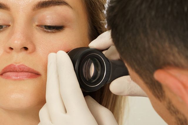
Non-melanoma skin cancers remain a very frequent reason for visits in dermatological offices. The most significant reason for their increased occurrence is an extensive exposure to UV light, however, aging of the population, immunosuppression or genetic background also take part in their pathogenesis. Both usually present as pink tumors or plaques and appear on sun-exposed skin.
The typical dermatoscopic features for Basal Cell Carcinoma (BCC) are known very well, and consist of linear branching vessels, blue clods, white, shiny blotches and strands, pink background, linear telangiectasia, and brown lines converging from a common base. Recently, data suggests trying to predict the histopathological subtype based on dermoscopic pattern which is also helpful in determining the appropriate treatment approach.
The described patterns for Squamous Cell Carcinoma (SCC) consist of white circles, white structureless areas, hairpin and glomerular vessels, white scales, keratin masses for well-differentiated tumors, and red background, polymorphous vascular pattern, and absence of white criteria in poorly differentiated malignancies. However, in some SCCs, setting the appropriate diagnosis might be difficult, and in some cases the features might overlap with other tumors.
Learning objectives:
- To summarize the known dermatoscopic features for BCC and SCC
- To categorize the dermatoscopic BCC patterns to predict the subtype before excision
- To underline some SCC characteristics to enhance the diagnostic efficacy of difficult lesions that might mimic other cancers