
Welcome letter from the SPC Chair
Prof. Lidia Rudnicka
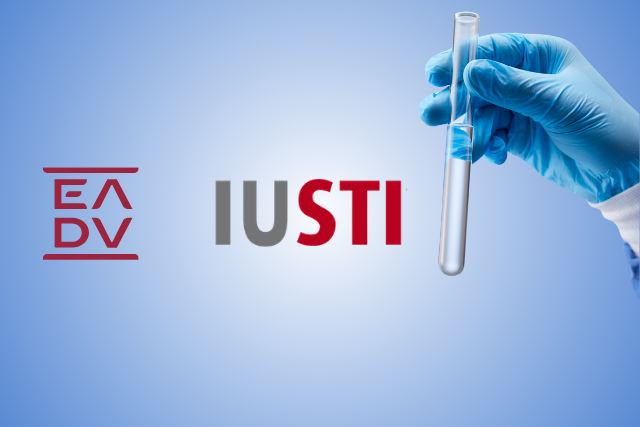
Joint EADV-IUSTI-EU Session on Venereology
Prof. Marinović and Prof. Tiplica chair a joint session by EADV and IUSTI on venereology: PCR in syphilis, neglected balanitis, and gonorrhea resistance.
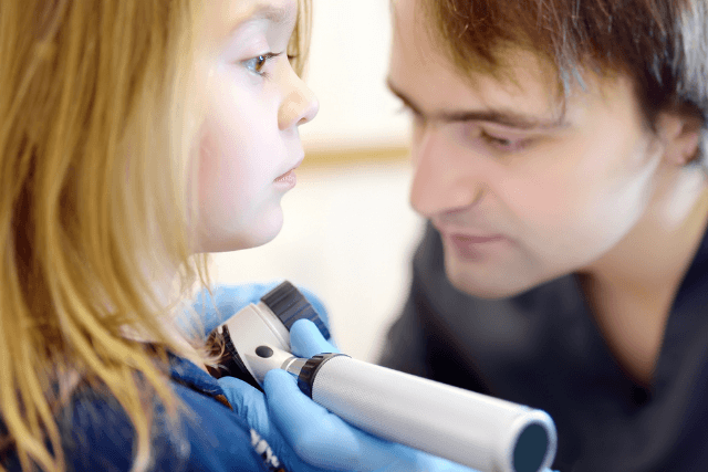
Focus on paediatric dermatology: Therapeutics
Discover the latest paediatric dermatology therapeutics, including off-label treatments, biologics, and JAK inhibitors for inflammatory and autoimmune skin diseases in children. Expert-led congress sessions focus on safe prescribing, innovative therapies, and improved patient outcomes.
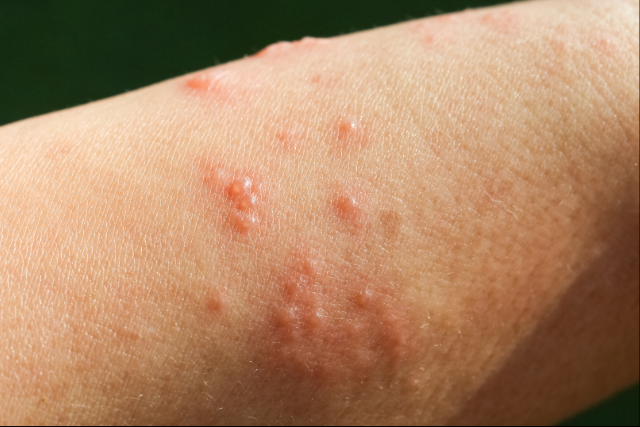
Bullous diseases
From underlying mechanisms and diagnostic approaches to emerging entities and therapeutic strategies, this session at the EADV Symposium 2026 aims to delivering a concise overview of the latest developments shaping clinical practice.
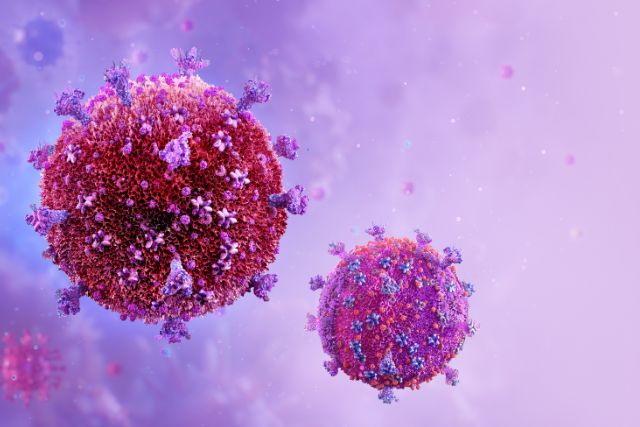
Prevention of STIs
This presentation will provide a clinically focused and evidence-based overview of PrEP and PEP, including indications, eligibility assessment, baseline investigations, follow-up protocols, and safety monitoring.
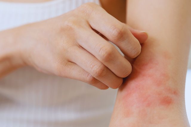
Atopic Dermatitis
This session explores the latest approaches to managing Atopic Dermatitis, including lifestyle measures, prevention strategies, and advances in systemic and biologic treatments. Experts will also discuss long-term safety data, emerging therapies, and how early, integrated care may help modify disease progression.

Pathogenesis and endotypes
Maciej Pastuszczak, Poland

Lifestyle, prevention and disease modification
Mette Sondergaard Deleuran, Denmark

Comorbidities: Management and workup
Stamatis Gregoriou, Greece
Treatment
Marie-Aleth Richard, France (Chair)
New vaccines
Sébastien Fouéré, France

PREP and PEP
Evdoxia Panou, Greece

Behavioural and social intervention
Valeska Padovese, Malta

Next generation diagnostics
Maciej Pastuszczak, Poland