
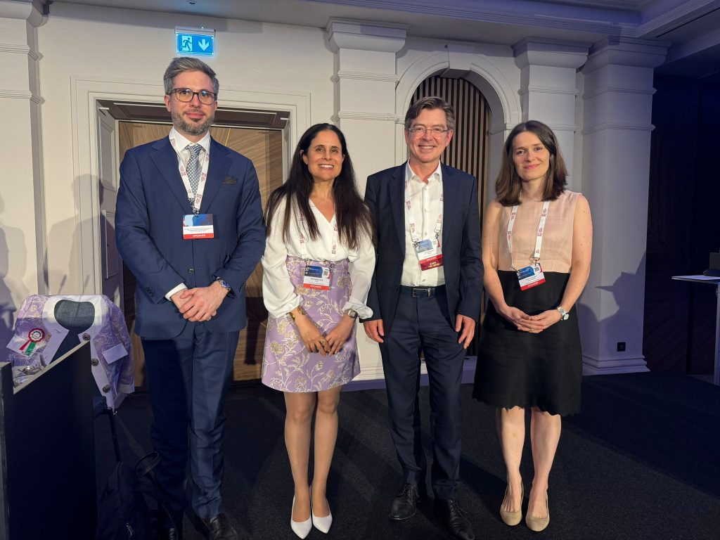
EADV Symposium 2024 Highlights
Welcome to the EADV Symposium 2024, taking place in the picturesque setting of St Julian’s, Malta. Catch a glimpse of each day’s action in this bite-sized round-up of scientific highlights.
Thursday
16 May 2024
Today's hot topics
What's new in: Acne
Presenting the latest the evidence on the management of acne
Acne is one of the most common diseases worldwide, not only in the adolescent post pubertal age group but also in the infantile and pre-pubertal age. Many myths still exist, and the importance of rational and evidence-based treatment is highlighted. This session presented the latest evidence on the management of acne with lasers and light therapy, dietary supplement use and the use of isotretinoin in order to facilitate rational decision making.
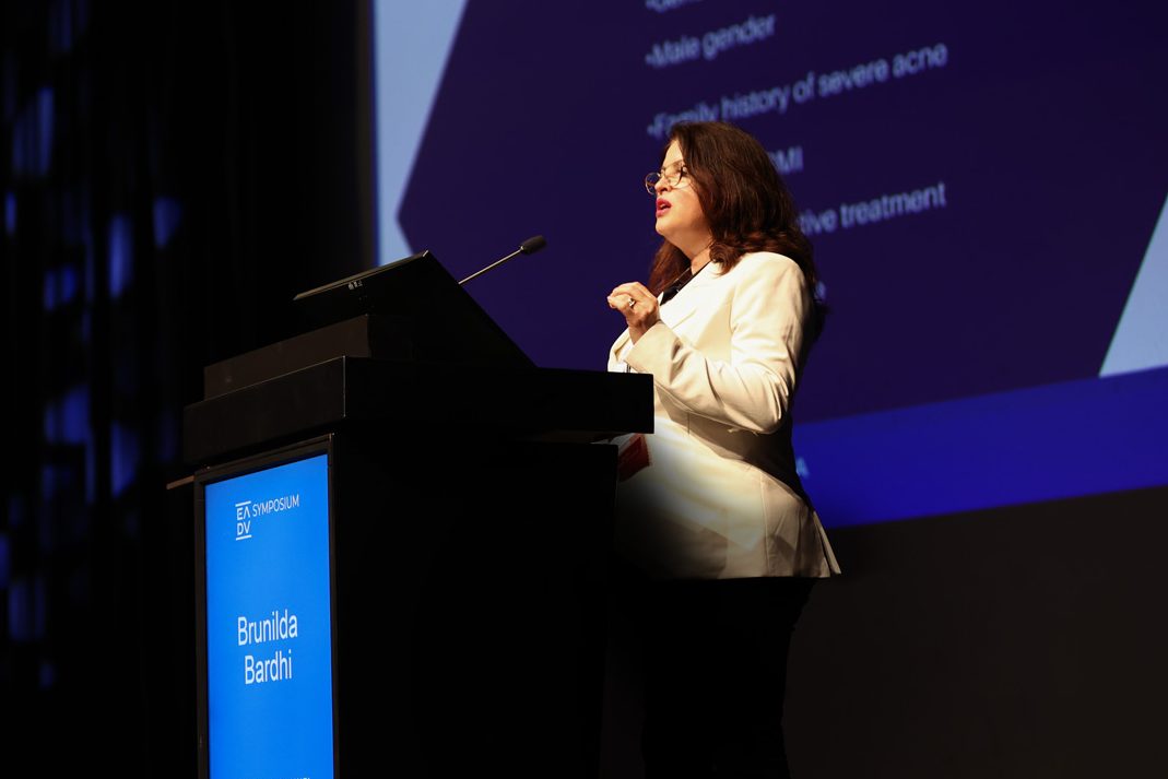
Laser and light therapy for acne and its scars
Dr. Brunilda Bardhi (Tirana, Albania) demonstrated how laser and light-based therapies alone and combined are efficacious methods for addressing the widespread problem of acne scars. These therapies target microbial colonisation, sebum production, and other underlying factors. For acne scars, non-ablative light, laser-based devices, and multi-modality treatments are demonstrating impressive results. However, complete removal of acne scars is not possible, and prevention remains key.
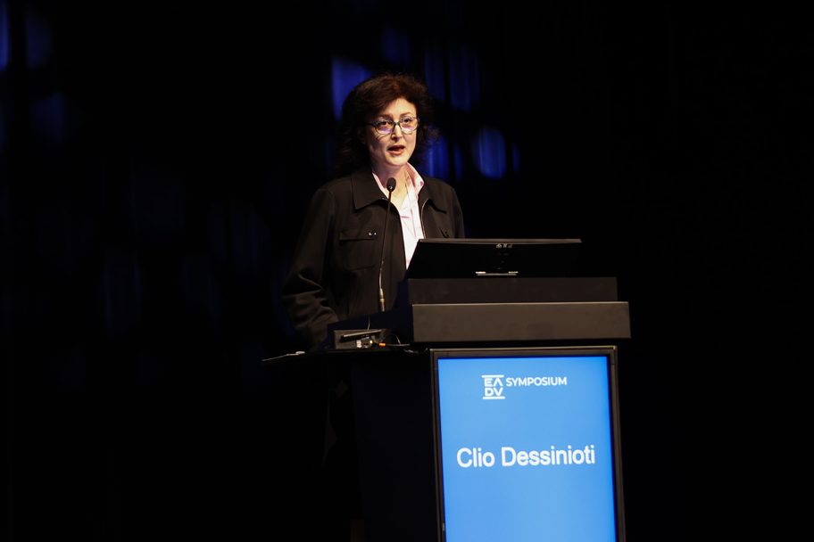
Navigating infantile and pre-pubertal acne
Dr. Clio Dessinioti (Athens, Greece) shed light on the distinct characteristics and complexities of infantile and prepubertal acne. The pivotal role of androgens, primarily dehydroepiandrosterone sulfate or ‘the acne hormone’, in the development of infantile acne was discussed. Although rare, infantile acne presents challenges with its heightened prevalence of inflammatory lesions and susceptibility to scarring, which can indicate underlying endocrine abnormalities. This was one of several examples of the importance of differential diagnosis. Given the nuances between acne across life stages, effective management necessitates a personalised approach that considers age, symptom severity, and hormonal imbalances.

The benefits and side-effects of isotretinoin
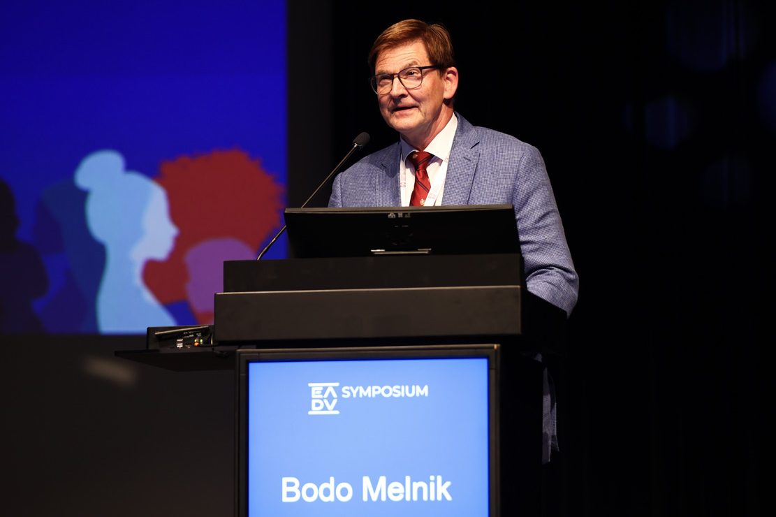
Through transcriptomics, Professor Bodo Melnik (Osnabrück, Germany) took symposium attendees on a 40-year journey into the growing understanding of isotretinoin. The beneficial genetic impact of isotretinoin is evident in its ability to upregulate p53, FoxO1, and FoxO3, promoting apoptosis of oil-producing sebocytes. However, isotretinoin also comes with potential side-effects, including dry skin and mucous membranes, pseudotumor cerebri, birth defects, depression, and very low-density lipoprotein hyperlipidaemia. While isotretinoin is effective in treating acne, understanding its genetic mechanisms can help doctors manage its use and potential side-effects more effectively. Further information can be found in Prof. Melnik’s recently published paper.
“The critical impact of isotretinoin is to reduce progenitor stem cells in the sebaceous glands, but this has not yet been proven and will be an exciting study opportunity for younger dermatologists.”
Piecing together the acne-diet puzzle
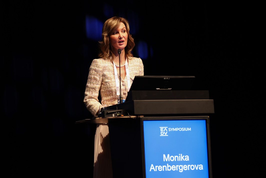
Prof. Monika Arenbergerova (Prague, Czech Republic) presented a captivating exploration of the link between the Western diet and acne. While there remains a lack of robust clinical trials, basic and translational research offers evidence of specific dietary factors that may influence the inflammatory cascade of acne. The strongest and most consistent evidence is of a link between acne and the consumption of milk and sugar. Glycaemic index (GI) is also a key player in the diet-acne relationship, impacting hormone levels that influence sebocytes and keratinocytes. Adiponectin, a protein secreted by adipose tissue, looks to be the next big area of exploration in acne pathogenesis. Adiponectin’s beneficial properties, including its inhibition of mTOR, underscore its potential significance in acne management.
“We believe that the hyperglycaemic diet, influenced by refined carbohydrates or milk, reduces levels of adiponectin, which has a direct connection to pro-inflammatory cytokines and to reduction of anti-inflammatory cytokines. This leads to sebaceous gland hyperplasia.”
Key takeaways
- With an increased understanding of the pathogenesis of acne, new and improved treatments are in use and on the horizon.
- Given the many underlying age-related, genetic, and dietary factors that can contribute to acne, a personalised approach to diagnosis and treatment is critical.
Focus on Atopic dermatitis: "Pathogenesis"
Exploring recent advances in atopic dermatitis
In recent years, there has been significant progress in our understanding of atopic dermatitis, including its pathophysiology, the characterisation of endotypes, and the development of novel treatments. This session aim provided an in-depth exploration of these advances, offering updates on the latest breakthroughs and crucial state-of-the-art knowledge for clinical practice.
The disease’s pathogenesis
Prof. Dr. Jan Gutermuth (Brussels, Belgium) discussed that atopic dermatitis (AD) is an inflammatory disease affecting the microbiome, chemical, physical, and immunological barriers, with systemic comorbidities, and highlighted the importance of identifying trigger factors (e.g. allergens) in certain patients. He noted that a multiple-marker approach would be useful to identify different disease endotypes and serological markers and added that, the pathogenesis of psychosocial factors such as stigmatisation is also important to avoid cumulative life course impairment, underlining a need for early treatment to restore health-related quality of life.
In the future, disease modification may represent a unique opportunity for intervention from the presymptomatic, innate immune response stages (with antigen presentation), to the adaptive immune response stage. He highlighted a need to identify relevant biomarkers to recognise those likely to develop atopic (e.g. food allergy, asthma, and rhinitis) or non-atopic (e.g. cardiovascular disease) comorbidities.
Atopic dermatitis: The significance of the microbiome
Dr Norbert Kiss, MD, PhD (Budapest, Hungary) discussed the relevance of the skin barrier and microbiome, and involvement of the gut in AD.
Dysbiosis of the skin is important in the development of the AD disease course. Staphylococcus aureus is more abundant in the skin of AD patients, particularly in lesions, and has an established pathogenisis. Different topical bacterio-therapueutic approaches show potential to normalise the skin microbiome.
The gut microbiome has an impact on AD and oral probiotic use (e.g. with Lactobacilli and Bifidobacteria,) during pregnancy, lactation and infancy may prvent AD development. However, oral prebiotics, synbiotics and postbiotics are not currently recommended owing from a lack of robust scienitifc evidence.
A warming world influences the cutaneous microbiome, and in turn, skin health
-
The latest on metabolomics
Dr. Helen Alexander (London, United Kingdom) discussed the links between the skin and gut microbiomes and host immune response in AD.
Skin is unique metabolic microenvironment, however, many studies of immunometabolism use lymphoid tissue or blood cells, rather than evaluating metabolite levels in the skin. Short-chain fatty acids (SCFAs) have anti-inflammatory effects, suppressing bacterial growth, while increased skin pH leads to increased protease activity, reduced lipid formation and inactivation of anti-microbial peptides promoting bacterial colonisation Glucose and folate metabolism may also mediate host interactions.
She discussed the importance of the gut microbiome, where imbalances in early life may affect immune system development, or in established AD, may induce inflammatory changes and reduce SCFA levels.
Harnessing metabolic interactions to treat AD include pre-, pro- and post-biotic AD therapy with exciting potential for the latter. However, improved understanding of the key mechanisms and quantification of metabolite levels (e.g. SCFA, glucose, and amino acids) are needed.
Examining environmental factors
In the final presentation, Prof. Roni Dodiuk-Gad (Haifa, Israel) highlighted the environmental factors affecting the disease course of AD based on her insights. These included irritants (e.g. hot water, scrubbing of skin, or clothes); microbial organisms (e.g. Staphylococcus aureus infection); stress; contact allergens (e.g. drugs, fragrance, metals, or rubber) and sweating; and aeroallergens (e.g. dust mites, animal dander, or pollen). Climate also affects the microbiome and dysregulation of the skin barrier.
She discussed how skin’s immune system protective mechanisms are involved in AD pathogenesis, and that environmental triggers induce type 2 inflammatory responses in the skin and upper and lower airways causing AD.
Prof. Dodiuk-Gad concluded by discussing climate change, declared by the WHO as “…the biggest health threat facing humanity”. Rising temperatures and extreme weather events, increasing atmospheric carbon dioxide with heat-trapping effects, boost a plant’s ability to produce pollen and extend the pollen season, which can change the skin microbiome causing a high incidence and prevalence of allergic disease.
Key takeaways
In the future, strategies may include barrier restoration for presymptomatic stages, with therapeutics to treat inflammatory cytokines in active AD, alongside possible concomitant use of other therapies.
The skin’s immune system protective mechanisms are involved in AD pathogenesis, and that environmental triggers induce type 2 inflammatory responses in the skin and upper and lower airways causing AD.
Plenary Session
Welcome to the EADV Spring Symposium
The EADV Spring Symposium holds a special place in our annual calendar, emphasising EADV’s commitment to inclusiveness in Europe while allowing for in-depth exploration of specific topics spanning the field of dermatology and venereology.
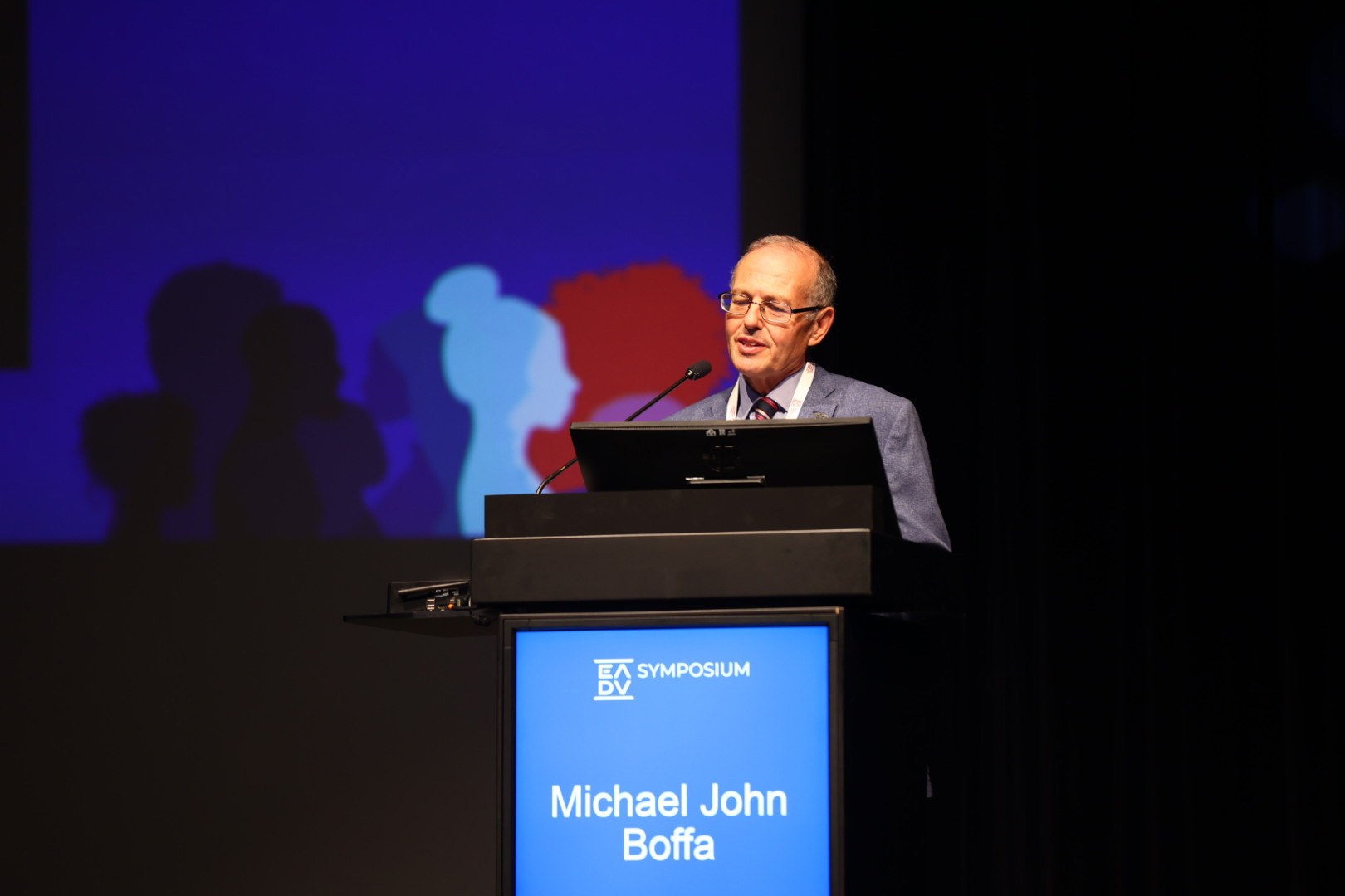
In today’s Plenary Session, Prof. Dr. Martin Röcken, EADV President, and Dr. Michael Boffa, welcomed delegates from 91 countries to this highly anticipated event, stressing the importance of Symposia for not only dermatologists but for patients too. “This symposium offers a unique opportunity to connect with specialists and colleagues from around the world who all share your same passion for dermatology and venereology. Over the next three days, we will experience a tailor-made programme covering valuable insights and the latest updates in crucial areas such as acne management, pigmentary disorders, atopic dermatitis, and paediatric dermatology.”
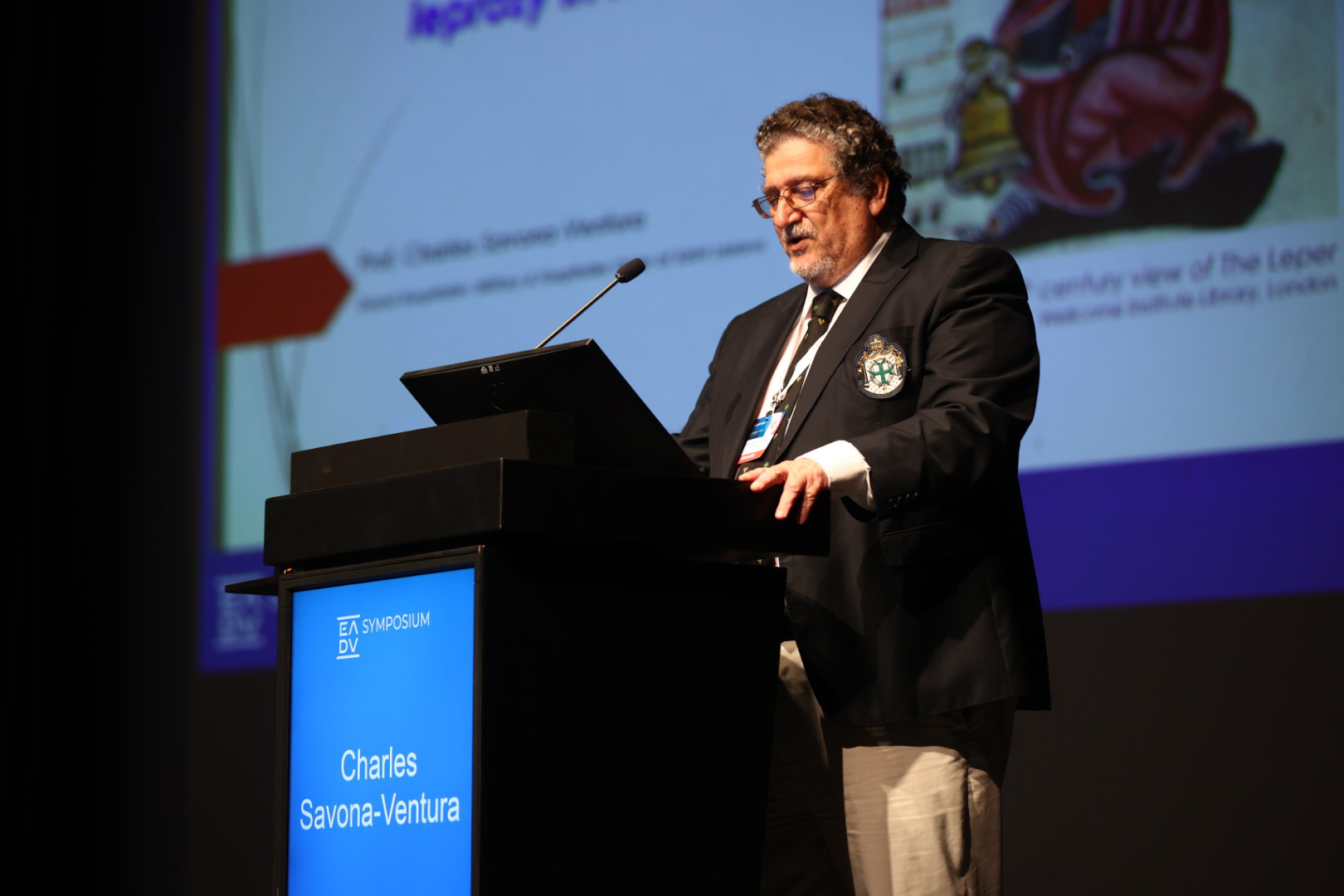
Within the session, Prof. Charles Savona-Ventura delivered a fascinating overview of the history of leprosy in Malta. The course of the disease and its prevalence was presented, taking attendees on a journey from the Bronze Age to recent decades, where Malta was declared free from indigenous leprosy. Throughout, Prof. Savona-Ventura discussed the lessons that have been learned on this immersive journey through time.
Despite historical attempts to segregate leprosy patients, such as placing restrictions on certain professions, these efforts proved ineffective. Launched in 1972, the Malta Leprosy Eradication Project was introduced to tackle cases within the country and, a decade later, multidrug therapy (MDT) was introduced as a tool capable of eradicating leprosy in a community. MDT, which uses a combination of three medicines (dapsone, clofazimine and rifampicin), rapidly became the standard treatment of leprosy. Since then, it has been supplied by World Health Organization free of charge to all endemic countries since 1995.
Friday
17 May 2024
Today's hot topics
What's New In: Melanoma
Exploring new discoveries in melanoma
It has been encouraging to see many discoveries and breakthroughs in recent years in the field of melanoma, many of which have significantly advanced our understanding of management and potential therapeutic options for patients. This session provided attendees on the latest insight on new imaging techniques, updates on safety margins and sentinel lymph node biopsy in melanoma, as well as adjuvant and neo-adjuvant therapy, and systemic treatment for advanced melanoma.
New imaging trends for diagnosis and follow-up
Prof. Mette Mogensen (Copenhagen, Denmark) delivered an enlightening talk on the potential of Photoacoustic Imaging (PAI), Reflective Confocal Microscopy (RCM), and Optical Coherence Tomography (OCT) to enhance diagnostic accuracy and monitoring—all at the bedside. These devices use lasers to create images through different methods, penetration depths, and resolutions. Prof. Mogensen showcased an image of malignant melanoma as displayed via PAI, which enables 3D molecular images through a handheld scanner, allowing clinicians to examine the lesion in real time. These impressive devices are likely to improve further with the addition of artificial intelligence.
Navigating safety margins and sentinel lymph node biopsy
Prof. Eduardo Nagore (Valencia, Spain) highlighted the challenges of Wide Local Excision (WLE) and the need for more precise techniques, such as Mohs surgery. He presented evidence that staged margin-control excision techniques, which are preferable for some cases of lentigo maligna and acral lentiginous melanomas, have resulted in lower recurrence rates than WLE. Prof. Nagore also discussed the debate around the value of sentinel lymph node biopsy (SLNB), expressing his opinion that it should be abandoned in cases where adjuvant therapy is already indicated. However, he believes SLNB should be offered to patients with a relatively high probability of modifying the management, such as those with clinical stage IIA.
Prevention and early diagnosis remain important, but remarkable progress has been made with immune checkpoint inhibitors and targeted therapies, with chemotherapy becoming much less important
Advances in adjuvant and neoadjuvant treatment
Prof. Petr Arenberger (Prague, Czech Republic) discussed advancements in adjuvant and neoadjuvant therapies for melanoma, both benefiting from immunotherapy. Specifically, anti-CTLA-4 and anti-PD-1 have shown efficacy in both malignant melanoma and the adjuvant setting, improving relapse-free survival. In neoadjuvant therapy, immunotherapy has outperformed targeted therapy. Furthermore, there has been a shift from inoperable tumours to targeting operable regionally advanced tumours (stages IIIB/C/D) without satellite and in-transit metastases, aiming for complete healing before surgery. Controversies and ongoing research aim to further refine adjuvant and neoadjuvant melanoma treatment.
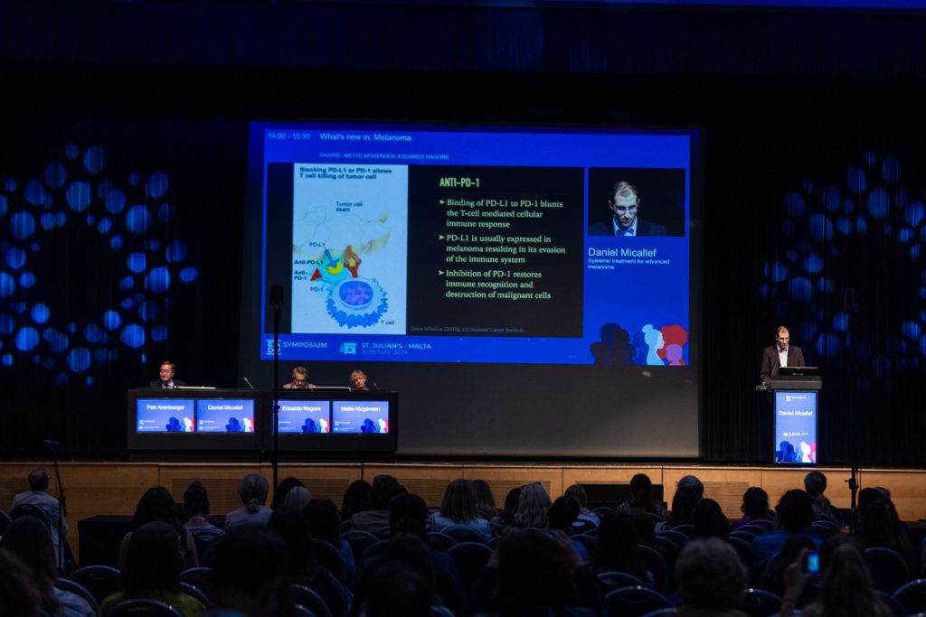
Game-changing systemic treatment for advanced melanoma
Dr. Daniel Micallef (Misda, Malta) presented the game-changing potential of immune checkpoint inhibitors (ICIs) and novel therapies for advanced melanoma. Highlighting the transformative impact of ICIs targeting CTLA-4, PD-1, and PD-L1, he discussed the enhanced efficacy of combination therapies for extending progression-free and overall survival. Notable advancements in treatment include the recent approval of nivolumab/relatlimab combination therapy and targeted therapy with BRAF-MEK inhibitors. Dr. Micallef underscored the importance of treatment sequencing and exploring triplet combinations of ICIs with BRAF and MEK inhibitors. Treatment of choice may depend on the existence of brain metastasis. Against this backdrop of innovation, a surge of cutting-edge clinical trials is propelling melanoma treatment to new frontiers.
Key takeaways
- New imaging techniques are enhancing the accuracy of melanoma detection and monitoring.
- Novel and targeted treatments across different settings and melanoma stages are increasing survival rates.
From clinical and dermatoscopic diagnosis to management
Cutting-edge updates on melanocytic lesions
Spitzoid lesions: The Schizophrenia of melanoma
Assoc. Prof. Aimilios Lallas (Thessaloniki, Greece) outlined the clinical recognition and management of benign Spitz nevi, offering compelling examples of the different clinical appearances of benign versus malignant Spitzoid. While dermatoscopy reveals certain diagnostic patterns, not all nevi conform to these patterns, reflecting the “schizophrenia” of Spitz nevi and the challenge of diagnosing malignancy. Given that 13% of Spitzoid lesions in individuals over 12 years are melanomas, with probability increasing with age, better methods of diagnosing malignant lesions are needed to inform management via excision or close monitoring.
What is ‘early’ melanoma?
Prof. Ana-Maria Forsea (Bucharest, Romania) highlighted the distinctions between “early” and “thin” melanoma, emphasizing that early melanoma does not equate to thin melanoma. It is, therefore, early detection of melanoma – thin or not – that is critical to better outcomes. There are challenges to dealing with early melanoma, however, including the key question of how early is early enough? Regardless of the answer, early detection through techniques like Total Body Photography and sequential dermatoscopy is critical for high-risk patients such as those with a family history of melanoma, past melanoma, or new growing lesions.
Dermatoscopic clues of melanotic melanoma
Prof. Danica Tiodorovic (Nis, Serbia) described the clinical presentation of amelanotic melanoma (AM), which accounts for 2-8% of melanomas and has a high misdiagnosis rate of 89%. Using case studies, Prof. Tiodorovic illustrated significant diagnostic challenges for AM, largely due to the lack of pigmentation and typical melanoma features. Importantly, she provided insights on dermatoscopic features of AM, with certain vascular patterns being key diagnostic indicators. Given the atypical presentation of AM and that it can mimic other conditions, awareness of the dermatoscopic clues of AM is critical to early detection and treatment.
Special techniques for special melanoma locations
Dr. Giulia Briatico (Naples, Italy) provided insights into melanoma occurring in “special locations” such as the face, acral regions, nails, and genitals. She highlighted the unique challenges of diagnosing these cases and the need for specialised diagnostic criteria. For example, the dermatoscopic inverse approach enhances diagnostic precision of lentigo maligna, while pigmentation patterns are crucial for differentiating between acral melanoma and acral nevus. Awareness and understanding of these unique characteristics can drive early detection and effective treatment.
“Spitz nevus believes he is melanoma. He behaves and dresses like melanoma. He grows like melanoma, in fact even faster, and sometimes even metastasises to the lymph nodes like melanoma. But, he is not melanoma. He can’t kill. Once he realises this, his life becomes meaningless, and he kills himself; he disappears.”
Key takeaways
- Melanoma comes in all shapes, sizes, and clinical presentations, necessitating diagnostic vigilance.
- Dermatoscopic identification of melanoma is especially crucial or those melanomas with atypical features or in atypical locations.
Focus on: Atopic dermatitis - Therapies
Therapeutic patient education and novel treatments for atopic dermatitis
This session focused on importance of therapeutic patient education, current topical and systemic treatments, and novel therapies for atopic dermatitis (AD).
In focus: Patients’ education
Through his involvement in setting up atopy schools and as scientific consultant on ‘Livros da Diana’ (books of a Portuguese child living with AD), Dr. Pedro Mendes-Bastos (Lisbon, Portugal) reiterated the importance patient education in consultations.
Therapeutic patient education (TPE) is recommended in European guidelines as a consensual transfer of skills from healthcare professionals to their patients. He noted that TPE should be evidence-based, and tailored to patient needs in an interdisciplinary setting. While multidisciplinary, well-structured, age-specific group programmes are key, the optimal format (e.g. face to face [F2F] vs digital, individual vs group) is to be determined.
A national survey in France showed that over 100 TPE tools are used, the majority were F2F with materials. However, use of online videos post-COVID-19 and WhatsApp may be attractive options to promote TPE. He concluded that while there are benefits of both F2F versus digital implementation, a sense of belonging to an AD community could be useful.
Education is crucial…we as doctors have to have the maximum of empathy…even in the time of JAK [inhibitors] and biologics
Investigating topical therapies
Assoc. Prof. Efstratios Vakirlis (Thessaloniki, Greece) gave an insightful overview of current knowledge on topical therapies and their use in current practice. He emphasised that baseline therapy (emollients, allergen avoidance and patient education) and topical treatment are indicated continuously in adults, regardless of AD severity.
Topical treatments remain the mainstay of therapy. Topical corticosteroids (TCs) are recommended first-line, while topical calcineurin inhibitors (TCIs) are approved for second-line use, supported by long-term safety. TCs and TCIs should be used for managing exacerbations and as proactive therapy for maintenance, and can be used with systemic agents (e.g. antimicrobials).
Over 30 novel topical compounds are in development for AD, including topical phosphodiesterase 4 inhibitors, janus kinase inhibitors (JAK), aryl hydrocarbon receptor agonists, transient receptor potential vanilloid 1 antagonists, and skin microbiome modulators. Greater understanding and novel products will help to improve AD care.
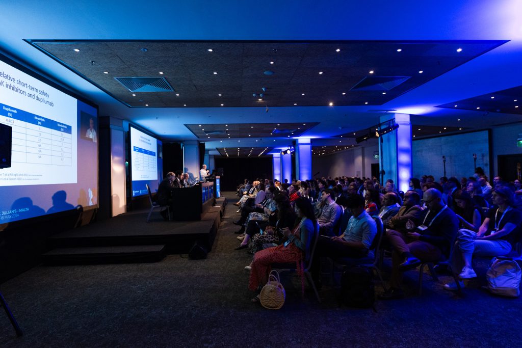
Biological agents or JAK inhibitors for moderate-to-severe AD
Prof. Carle Paul (Toulouse, France) sought to guide when to use specific treatments in AD, a market dominated by biological and topical agents.
Biologics for AD include anti-IL-14α, anti-IL-13, and anti-IL31 inhibitors, and JAK 1, 2 inhibitors, each with different dosing schedules. He cautioned that in vitro selectivity does not always correlate with in vitro data.
In randomised clinical trials, short-term comparisons of JAK inhibitors (abrocitinib and upadacitinib) showed greater efficacy in the proportions of patients achieving EASI 90, with a rapid effect on pruritus, versus dupilumab. Differences in efficacy reduced over time.
When stopping treatment, relapse of disease can be more gradual with dupilumab than JAK inhibitors. Also, while the incidence of conjunctivitis is higher with dupilumab, acne, folliculitis, herpes zoster, and upper respiratory tract infections are higher with JAK inhibitors, with dose-effects from long-term use.
Upcoming aspects of management of atopic eczema
Prof. Dr. Johannes Ring (Munich, Germany) noted that allergies and AD are a global problem, with hygiene appearing to be the most popular hypothesis together with parasites, pollutants, and climate change. He then focused on the growing incidence of AD and the role of the brain and psyche.
Interaction between the environment and organism employs both the non-structured immune system and structured nervous systems, with the itch/scratch response occurring in the brain. Functional MRI shows that when itch is induced in non-lesional skin, there is a state of active downregulation, while in lesional skin it is upregulated. Psychological conditioning is also relevant in allergy and may involve psychosomatic interference with histamine release. The brain, skin, and immune system are also closely connected.
Prof. Ring concluded by underlining that physicians should be aware of therapeutic strategies from broad to more targeted options and discussed several therapeutic agents. He noted, however, that not all drugs would solve all problems because of non-responders, adverse effects, and different treatment durations before echoing that patient education and empathy, alongside long-lasting remission and improvement, are crucial.
Key takeaways
- Therapeutic education with a multidisciplinary approach is key to improving patient involvement and adherence.
- Background therapies should be used concurrently with topical therapies, with the choice of specific therapeutic agent being used where it is most appropriate.
Saturday
18 May 2024
Today's Hot topics
What's New In: Hair disorders
Hair disorders, alopecia, and the potential for AI
Today marks the third and final day of the EADV Symposium 2024. Today’s programme showcased four ‘What’s new in:’ sessions to explore hair disorders, pruritis and prurigo, paediatric dermatology, and bullous diseases. Here’s a glimpse of the action from this morning’s hair disorders session.
Paediatric hair disorders
Dr. Liam Mercieca (Msida, Malta) started the session by recapping some of the most common hair disorders in children.
He presented a systematic approach to diagnosing paediatric hair loss, by determining if the cause is congenital (diffuse or focal) or acquired (scarring or non-scarring), and highlighted that disease is not always immediately apparent, as approximately 50% of hair loss is required to see the scalp.
Transient neonatal hair loss, thought to be due to sleep pressure, is caused by factors in pregnancy and hair shedding in utero. It is not recommended to change sleeping position because of cot death risk, and hair always grows back.
Underlying psychological causes should be considered for hair loss due to behavioural issues (e.g. trichotillomania), with counselling to stop hair pulling. Paediatric hair loss should aways be taken seriously, with early diagnosis and treatment initiation, particularly scarring alopecias and tinea capitis to avoid permanent damage.
There is a continuous need for human expertise. Professionals should always oversee AI systems and make the final decision on patient care

A new therapeutic landscape for alopecia areata
Dr. Francesco Tassone (Rome, Italy) reminded the attendees that up until 2023 there were no approved therapies for alopecia areata (AA), an autoimmune chronic disease with an unpredictable evolution, relapses and the worst prognosis for paediatric hair disease. Topical corticosteroids and immunotherapies, recommended as first-line therapy, are associated with efficacy and safety issues.
He highlighted that the AA therapeutic landscape has changed completely in the last two years, with many new compounds in development. Notably, JAK inhibitors (baricitinib and ritlecitinib) have been approved and are pending approval (deuruxolitinib) for patients aged > 12 years with AA, while other drugs approved for different indications are used off-label. Phase 3 trials of JAK inhibitors and follow up data show promising efficacy and safety results, with high proportions of hair regrowth maintained over time.
Infections and hair loss
Prof. Giuseppe Micali (Catania, Italy) classified hair loss into three main groups.
Scalp infections that may cause hair loss include tinea capitis, associated with broken hair (not loss) occur mostly in children with varying epidemiology, and are non-scarring. Fungal culture is the gold standard for diagnosis, with immediate treatment and identification of common trichoscopy features. Rare forms (favus and kerion celsi) require prompt treatment to avoid scarring and hair loss.
Systemic infections that may cause hair loss include secondary syphilis and viral infections. Symptomatic or essential syphilitic alopecia has distinct clinical patterns associated with systemic, mucosal or cutaneous manifestations. COVID-19 alopecia has been associated with telogen effluvium, which is more noticeable in females, and may increase AA worsening but is not a cause of new diagnoses.
Scalp infections with a controversial role in hair loss include folliculitis decalvans, a rare form of progressive neutrophil-associated scarring alopecia, often thought to be related to a specific microbiome or microbiological signature. It is characterised by tufted hair folliculitis caused by fibrous tissue contraction and topical or systemic antibiotics remain the treatment of choice.
AI in dermatology – are we ready?
In the final engaging presentation, Prof. Dr. Alexander Katoulis (Athens, Greece) shared the potential for artificial intelligence use (AI) in medicine to revolutionise how diseases are diagnosed and treated, particularly in image-based fields such as Dermatology.
Trichoscopy, the most useful diagnostic tool for hair disorders, notably for alopecia, may facilitate AI application. Early algorithms required short hair for evaluation; however, newer systems can analyse hair growth and rate, density and diameter and generate anagen/telogen ratio and digital trichogram data, without the need for hair trimming. However, current developments have not yet integrated its core (i.e. trichoscopic diagnostic clues and patterns).
Challenges in AI-assisted imaging include image quality and standardization, and the presence of artifacts or anatomic sites. Limitations include variations in hair characteristics, accuracy and reliability, data quality and bias.
Prof. Katoulis remarked it was important for physicians to take an active role in AI tool development to facilitate their effective implementation in clinical practice, where AI should be used to enhance, not replace, the physician’s abilities and the physician-patient relationship.
Key takeaways
- The introduction of JAK inhibitors for patients over 12 with alopecia areata show promising efficacy and safety, marking a substantial improvement over traditional therapies.
- Despite AI advancements, AI should support, not replace, physicians in clinical practice.
Breaking news
Spring Symposium: The forefront of dermatology innovation
To conclude this year’s Spring Symposium, three ‘Breaking News’ sessions featured within Saturday’s programme, where the latest advances in treating of a wide range of dermatological conditions were discussed. The final of the three explored the future of dermatology and presented key topics that promise to shape clinical practice in the years to come.
This included precision medicine, which holds the potential to enhance diagnostic precision and guide individual therapeutic decisions, and artificial intelligence, which is poised to revolutionise and streamline diagnoses.
Expression profiling: The present and future of inflammatory skin diseases
Prof. Michel Gilliet (Lausanne, Switzerland) offered a fascinating talk on the need for skin disease classification to advance from morphological to molecular classification. He highlighted the revolutionary potential of therapies targeting molecular events in dermatology, noting that response variability suggests wrong diagnoses or that the right molecular pathways are not being targeted. Prof Gilliet shared an individualised molecular profiling approach adopted within his clinic using Nanostring technology, which has proven effective in diagnosing inflammatory skin conditions, enabling tailored therapies, and enhancing precision medicine.
In our clinic, Nanostring technology is fully implemented and we are looking at all inflammatory disease with this molecular approach. We are now trying to develop a network across Europe that will do this with us. I am convinced expression profiling of skin diseases is the present and certainly the future.
Somatic mutations in inflammatory diseases
Prof. Yuval Ramot (Jerusalem, Israel) described the intricate world of somatic mutations, which are increasingly being linked to inflammatory cutaneous diseases. Through four case studies, Prof. Ramot illustrated the impact of somatic mutations on diseases with skin involvement, such as VEXAS syndrome – a newly discovered disorder connecting previously unrelated inflammatory syndromes and serving as a prototype for a new class of haemato-inflammatory diseases. Advancements in mutation identification and tracking promise to define new disease syndromes, uncover novel biomarkers, and pave the way for precision medicine.
Apps and AI: Friends or foes?
Dr. Tobias Sangers (Rotterdam, Netherlands) explored the potential benefits and drawbacks of AI-assisted dermatology smartphone apps. These apps can increase skin cancer awareness, promote protective behaviours, and even perform on par with dermatologists and better than junior doctors in diagnosing pigmented skin cancer. However, AI remains on the scales – a friend that increases skin cancer awareness and diagnosis and an enemy that produces false positives and missed diagnoses. The EADV Task Force has a position statement on the use of these technologies within dermatology, recognising its benefits for patients and offering recommendations to drive its safe and effective use.
New and emerging therapies for melanoma
Prof. Martin Röcken (Tübingen, Germany) outlined the dynamic landscape of melanoma treatment. While conventional treatments remain crucial, personalised and combination therapies are enhancing survival rates and minimising adverse effects. These include anti-PD1 mAbs, combined BRAF/MEK inhibitors, anti-PD-1/anti-CTLA-4 mAb combinations, and anti-LAG3 targets. Ongoing research is also exploring biomarker-based approaches, aiming to identify resistance-associated genes. Prof. Röcken finished his talk by emphasising the potential of combining immune therapy with novel targets that still need to be identified.
Key takeaways:
- With a growing understanding of molecular pathways and somatic mutations, precision medicine is gaining momentum.
- AI is changing self-diagnosis and may be set to change clinical diagnosis, necessitating dermatologists to keep abreast of technological advancements.
- New melanoma treatments, including personalised and combination therapies, are improving survival rates and reducing side-effects.